
Diploma Dental Assistant (DDA) Course 2 Years. Mobile Number-01987073965,01797522136.This Course Contains 18 Subject. There are Total 4 Semester. The 1st Semester Contains 5 Subject Which are Human Anatomy & Physiology, Chemistry & Pharmacology, Study of OTC, First Aid & Practice Of Medicine, Hematology & Pathology. 2nd Semester Contains 5 Subject Which are Dental Anatomy & Physiology, Anatomy of Headneck, Neuro Anatomy & Trigeminal Nerves, Dental Filling Materials, Clinical Practice In Dental Surgery.3rd Semester Contains 4 Subject And The Last Semester also Contains 4 Subject.

Location of Diploma Dental Assistant Course 2 Years
Location of Diploma Dental Assistant Course 2 Years. MobileNumber.01987073965,01797522136. HRTD Medical Institute , Abdul Ali Madbor Mention, Section-6, Block-Kha, Road-1, Plot-11, Mirpur-10 (Gol-Chattar) Metro Rail Pilar NO-249, Dhaka-1216. It is situated by the West Side of Agrani Bank, the South Side of Fire Service, Islami Bank, Janata Bank, Social Islami Bank, Medinova, Ibrahim Diabetic Hospital, the North Side of Baitul Mamur Jame Mosjid, Grave of Baitul Mamur Jame Mosjid, and East Side of Maliha Apartment.
Course Fee For Diploma Dental Assistant Course 2 Years
Admission Fee=16,500/-,Monthly Fee 3000×24=72,000/-,Exam Fee=1000×4=4,000/-, Total Course Fee=92,500/-.Books Fee for Every Semester 1,500/-.
Diploma Dental Assistant Course 2 Years Admission Eligibility
Diploma Dental Assistant Course 2 Years Admission Eligibility. Mobile Number. 01987073965. 01941123488, 01797522136. SSC or Equivalent/HSC/ Degree/ Masters from any Background (Science/ Arts/ Commerce/ Technical).
Documents for Admission in Diploma Dental Assistant Course 2 Years
Diploma Dental Assistant Course 2 Years Course in Dhaka. Mobile No: 01987-073965, 01797-522136. HRTD Medical Institute. Document Needed: Photocopy of Certificate, Photocopy of NID, Passport Size Photo 4 Pcs. Without NID, a Birth Certificate is allowed for an emergency case.
Teachers For Diploma Dental Assistant Course 2 Years
- Dr. Sanjana Binte Ahmed, BDS, MPH, Assistant Course Director
- Dr. Nazmun Nahar Juthi, BDS, PGT
- Dr. Md. Sakulur Rahman, MBBS, CCD (BIRDEM), Course Director
- Dr. Suhana, MBBS, PGT Medicine
- Dr. Kamrunnahar Keya, BDS, PGT (Dhaka Dental College)
- Dr. Danial Hoque, MBBS, C-Card
- Dr. Tisha, MBBS, PGT Gyne, Assistant Course Director
- Dr. Afrin Jahan, MBBS, PGT Medicine
- Dr. Ananna, MBBS
- Dr. Lamia Afroze, MBBS
- Dr. Amena Afroze Anu, MBBS, PGT Gyne, Assistant Course Director
- Dr. Farhana Antara, MBBS,
- Dr. Farhana Sharna, MBBS
- Dr. Bushra, MBBS
- Dr. Turzo, MBBS
- Dr. Shamima, MBBS, PGT Gyne
- Dr. Alamin, MBBS
- Dr. Benzir Belal, MBBS
- Dr. Disha, MBBS
- Dr. Mahinul Islam, MBBS
- Dr. Tisha, MBBS, PGT Medicine
- Dr. Anika, MBBS, PGT
- Dr. Jannatul Ferdous, MBBS, PGT Gyne
- Dr. Jannatul Aman, MBBS, PGT
- Dr. Rayhan, BPT
- Dr. Abu Hurayra, BPT
- Dr. Sharmin Ankhi, MBBS, PGT Medicine
- Md. Monir Hossain, B Pharm, M Pharm
- Md. Monirul Islam, B Pharm, M Pharm
- Md. Feroj Ahmed, BSc Pathology, PDT Medicine
Practical Classes For Diploma Dental Assistant Course 2 Years
- Heart Beat, Heart Rate
- Heart Sound,Pulse
- Blood Pressure, Hypertension, Hypotension
- First Aid Box
- Auscultation
- Inhaler, Rotahaler
- Nebulizer
- Glucometer Blood Glucose
- Injection I/V
- Injection I/M
- Cleaning,Dressing,Bandaging
- Saline
- CPR
- Stitch
- Body Temperature
- Nasal Tube Gel ,Hand Wash
- Blood Grouping
- Cyanosis, Dehydration Test, Edema Test
Subjects For Diploma Dental Assistant Course 2 Years
Diploma Dental Assistant Course 2 Years .This Course Contains 18 Subject in 4 Semester. Mobile Number: 01987073965,01797-522136
1st Semester Subjects
- Human Anatomy & Physiology-1
- Pharmacology-1
- Study of OTC Drugs
- First Aid-1 & 2 & Practice of Medicine
- Hematology & Pathology for Medical Practice
2nd Semester Subjects
- Dental Anatomy & Physiology
- Anatomy of Headneck
- Neuro Anatomy & Trigeminal Nerves
- Dental Filling Materials
- Clinical Practice In Dental Surgery
3rd Semester Subjects
- Dental Abscess & Treatment
- Community Medicine
- Oral Surgery & Sterilization
- Conservative Dentistry & Radiology
4th Semester Subjects
- Dental Pharmacology 1 & 2
- Oral Abscess & Treatment
- Dental Emergency
- Dental Surgery & Prosthetics

HRTD Medical Institute Mirpur-10,Golchattar 01797-522136,01987-073965
Practical Class for Diploma Dental Assistant Course 2 Years
- Blood Pressure
- Hypertension & Hypotension
- Heart beat ,Heart rate, Heart Sound, Pulse
- Tooth Anatomy
- Headneck Anatomy
- Instrument Identify
- Scaling
- Diagnosis Procedure
- Filling Materials
- Root Canal Treatment
- Canal Shaping
- Obturation Sealing
- Children root canal treatment
- X-ray taking Procedure
- Crown Preparation for cap
- RPD Making (denture)
- Stitching
- Extraction Procedure
- Impress & Model making
- First Aid Box
- Auscultation
- Inhaler,Rotahaler
- Diabetes
- Injection I/V & I/M
Some Practical Class Details Given For Diploma Dental Assistant Course 2 Years
Scaling
Dental scaling is a professional cleaning procedure to remove plaque and hardened tartar (calculus) from tooth surfaces, preventing gum disease (gingivitis/periodontitis) by cleaning above and sometimes below the gumline, often paired with polishing for smoothness, using ultrasonic tools or manual instruments for a deep clean, ensuring healthier gums and fresher breath.
What it is:
- Plaque & Calculus Removal: Removes sticky bacterial film (plaque) and its hardened form (calculus/tartar) that brushing can’t eliminate.
- Gum Disease Treatment: A key non-surgical treatment for gum disease (periodontitis).
- Deep Cleaning: For deeper issues, it’s part of scaling and root planing (SRP), cleaning below the gumline where bacteria hide.
How it’s done:
- Tools: Dentists use ultrasonic scalers (vibrating tips with water spray) and/or manual instruments.
- Techniques:
- Supragingival: Above the gumline (regular cleaning).
- Subgingival: Below the gumline (deep cleaning).
- Polishing: Often follows to smooth teeth and remove stains for a clean finish.
- Comfort: Local anesthesia or numbing gel can be used for sensitive areas.
Why it’s important:
- Prevents Disease: Stops inflammation, bleeding gums, and tooth loss.
- Aesthetics: Removes stains for whiter teeth and fresher breath.
- Health: Keeps your mouth healthy, especially if you have gum pockets over 3mm deep.
Aftercare:
- Expect tenderness or slight bleeding for a few days.
- Maintain strict daily brushing and flossing, plus regular dental visits (every 3-6 months), to prevent buildup from returning.
Polishing
Dental polishing smooths tooth surfaces, removing stains, plaque, and debris to leave teeth shiny, feel clean, and prevent bacteria buildup, often done after scaling (deep cleaning) with an abrasive paste and a rotating rubber cup or air/water jet, improving both oral hygiene and smile aesthetics.
What it is & How it works
- A Finishing Touch: It’s the final step in a professional cleaning, making teeth glossy and resistant to new plaque.
- Tools: A slow-speed drill with a rubber cup (prophy cup) or an air polisher (using water, air, and powder like baking soda) is used.
- Process: A gritty polishing paste is applied as the cup or air jet scrubs the teeth to remove stains from coffee, tea, wine, and smoking.
Key Benefits
- Smoother Surface: Makes teeth feel slick and clean.
- Stain Removal: Eliminates extrinsic (surface) stains for a brighter appearance (not true whitening).
- Plaque Control: A smoother surface makes it harder for plaque to stick.
- Fresher Breath: Removes odor-causing bacteria.
Types of Polishing
- Rubber Cup Polishing: Most common, uses a cup with abrasive paste.
- Air Powder Polishing: Uses pressurized water, air, and powder for efficient stain removal, great for hard-to-reach areas.
Is it always needed?
- Selective Polishing: Dentists often choose to polish only when needed (e.g., for heavy stains or specific needs) rather than as a routine for everyone, to prevent enamel wear.
Some Subjects Details Given For Diploma Dental Assistant Course 2 Years
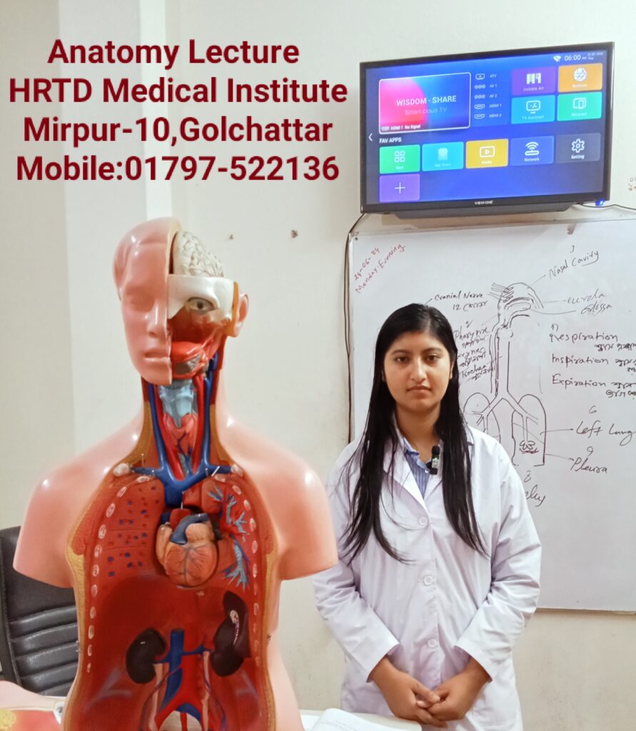
Human Anatomy & Physiology for Diploma Dental Assistant Course 2 Years
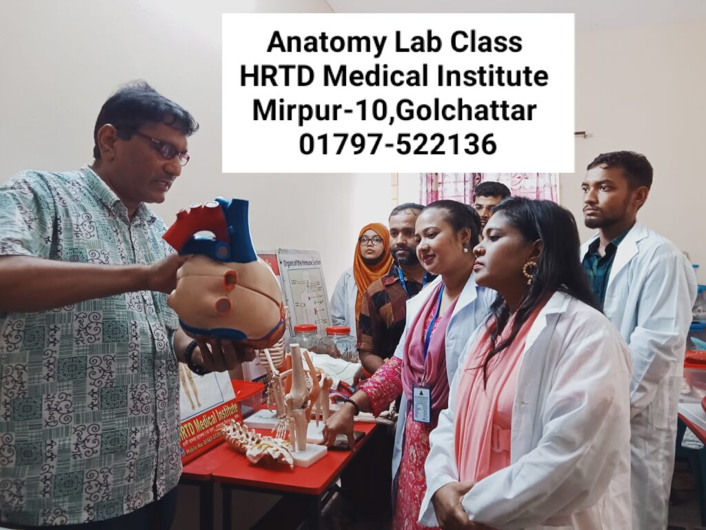
The Study of the body Structure and its function is Anatomy & Physiology. Here we discuss the systems of the human body and its organ, Tissues, and cells. The systems of the human body are the digestive system, Respiratory system, Cardiovascular system, Skeletal system, Muscular system, nervous system, Endocrine system, Immune System, Integumentary System and Urinary System.
1. Anatomy
Definition
Anatomy is the branch of science that studies the structure of the human body and the relationship between body parts.
Anatomy deals with “what the body is made of and where the parts are.”
Branches of Anatomy
- Gross (Macroscopic) Anatomy
- Study of structures visible to naked eye
- Example: heart, lungs, bones
- Microscopic Anatomy
- Study of structures seen under microscope
- Includes:
- Histology – study of tissues
- Cytology – study of cells
- Developmental Anatomy
- Study of growth from fertilization to adulthood
- Includes embryology
- Regional Anatomy
- Study of specific regions (head & neck, thorax)
- Systemic Anatomy
- Study of body systems (digestive, respiratory, etc.)
- Clinical Anatomy
- Application of anatomy in diagnosis and treatment
2. Physiology
Definition
Physiology is the branch of science that studies the functions of the human body and how body parts work.
Physiology deals with “how the body works.”
Branches of Physiology
- Cell Physiology
- Systemic Physiology
- Cardiovascular physiology
- Respiratory physiology
- Renal physiology
- Nervous system physiology
- Neurophysiology
- Endocrine Physiology
- Pathophysiology
- Study of functional changes in disease
3. Levels of Organization of the Human Body
- Chemical level (atoms, molecules)
- Cellular level
- Tissue level
- Organ level
- System level
- Organism level
4. Basic Tissues of the Body
- Epithelial tissue
- Connective tissue
- Muscle tissue
- Nervous tissue
5. Major Systems of the Human Body
| System | Main Function |
|---|---|
| Skeletal | Support & protection |
| Muscular | Movement |
| Nervous | Control & coordination |
| Endocrine | Hormonal regulation |
| Cardiovascular | Blood circulation |
| Respiratory | Gas exchange |
| Digestive | Nutrition & digestion |
| Urinary | Waste excretion |
| Reproductive | Reproduction |
| Integumentary | Protection & temperature control |
6. Relationship Between Anatomy & Physiology
| Anatomy | Physiology |
|---|---|
| Structure | Function |
| Form | Action |
| Static study | Dynamic study |
কয়েকটি সিস্টেম সম্পর্কে আলোচনা করা হলো:
1. Skeletal System
Function:
- Gives shape and support to the body
- Protects vital organs (brain, heart, lungs)
- Helps in body movement
- Produces blood cells in bone marrow
Main Parts:
- Bones (206 bones)
- Joints
- Cartilage
2. Muscular System
Function:
- Enables movement of the body
- Maintains posture
- Produces heat
Types of Muscles:
- Skeletal muscle – voluntary
- Smooth muscle – involuntary
- Cardiac muscle – found in the heart
3. Nervous System
Function:
- Controls and coordinates body activities
- Receives and responds to stimuli
- Responsible for thinking, memory, and emotions
Main Parts:
- Brain
- Spinal cord
- Nerves
4. Cardiovascular System
Function:
- Circulates blood throughout the body
- Transports oxygen, nutrients, and hormones
- Removes waste products
Main Parts:
- Heart
- Blood
- Blood vessels (arteries, veins, capillaries)
5. Respiratory System
Function:
- Helps in breathing
- Supplies oxygen to the blood
- Removes carbon dioxide from the body
Main Parts:
- Nose
- Trachea
- Lungs
- Alveoli
6. Digestive System
Function:
- Digestion of food
- Absorption of nutrients
- Elimination of waste
Main Parts:
- Mouth
- Esophagus
- Stomach
- Intestines
- Liver and pancreas
Pharmacology-1 for Diploma Dental Assistant Course 2 Years
1. Definition of Pharmacology
Pharmacology is the branch of medical science that deals with drugs, their sources, actions, uses, side effects, and mechanisms in the human body.
2. Drug – Definition
A drug is a chemical substance that is used to diagnose, prevent, treat, or cure disease.
3. Branches of Pharmacology
- Pharmacokinetics – What the body does to the drug
- Pharmacodynamics – What the drug does to the body
- Pharmacotherapeutics – Use of drugs in treatment
- Toxicology – Study of harmful effects of drugs
- Chemotherapy – Drugs used to treat infections and cancer
4. Pharmacokinetics (ADME)
A – Absorption:
How a drug enters the bloodstream
D – Distribution:
How the drug spreads in the body
M – Metabolism:
Breakdown of drugs mainly in the liver
E – Excretion:
Removal of drugs mainly through kidneys (urine)
5. Pharmacodynamics
- Drug action and effect
- Receptor interaction
- Dose–response relationship
Example:
Paracetamol reduces pain and fever.
6. Routes of Drug Administration
- Oral (by mouth)
- Sublingual
- Intravenous (IV)
- Intramuscular (IM)
- Subcutaneous (SC)
- Topical
- Inhalation
7. Types of Drugs
- Analgesics (pain killers)
- Antibiotics
- Antipyretics (reduce fever)
- Antiseptics
- Sedatives
8. Adverse Drug Reactions (ADR)
- Nausea
- Vomiting
- Allergy
- Drowsiness
9. Importance of Pharmacology for Nurses / Physiotherapists
- Safe drug administration
- Understanding drug effects
- Prevention of medication errors
- Patient education
10. Common Terms
- Dose: Amount of drug given
- Therapeutic effect: Desired effect
- Side effect: Unwanted effect
- Overdose: Excess amount of drug
Study of OTC Drugs for Diploma Dental Assistant Course 2 Years
OTC Drugs are important for all Medical Assistant Courses. These Drugs are Emergency and safe for the patients. The Study of OTC Drugs improves the quality of practice. Some OTC Drugs are Albendazole, Ascorbic Acid, Calcium, Multivitamins, Vitamin B Complex, Omeprazole, Oral Rehydration Salt, Salbutamol etc.
Definition
OTC drugs are medicines that can be purchased without a doctor’s prescription and are used for minor and self-limiting illnesses.
Purpose of OTC Drugs
- Relief of common symptoms
- Easy access to treatment
- Cost-effective healthcare
- Reduces burden on hospitals
Characteristics of OTC Drugs
- Safe when used as directed
- Low risk of serious side effects
- Clear labeling and instructions
- Suitable for self-medication
Classification of OTC Drugs
1. Analgesics & Antipyretics
Used for pain and fever.
| Drug | Use |
|---|---|
| Paracetamol | Fever, mild pain |
| Ibuprofen | Pain, inflammation |
2. Antacids
Used for acidity and indigestion.
| Drug | Use |
|---|---|
| Aluminum hydroxide | Neutralizes acid |
| Magnesium hydroxide | Relieves acidity |
3. Cough & Cold Preparations
- Antitussives – suppress cough
- Expectorants – remove mucus
- Decongestants – relieve nasal block
Examples:
- Dextromethorphan
- Guaifenesin
- Phenylephrine
4. Antihistamines
Used for allergy.
| Drug | Use |
|---|---|
| Cetirizine | Allergic rhinitis |
| Loratadine | Urticaria |
5. Antidiarrheal Drugs
Used for diarrhea.
| Drug | Use |
|---|---|
| ORS | Prevents dehydration |
| Loperamide | Reduces bowel movement |
6. Laxatives
Used for constipation.
- Bulk-forming: Isabgol
- Osmotic: Lactulose
- Stool softeners
7. Topical Preparations
Used on skin.
- Antiseptics (Povidone-iodine)
- Antifungal creams
- Pain relief gels
8. Vitamins & Minerals
- Vitamin B-complex
- Vitamin C
- Iron supplements
Dosage Forms of OTC Drugs
- Tablets
- Syrups
- Capsules
- Creams & ointments
- Drops
- Powders
Advantages of OTC Drugs
- Easily available
- Saves time & money
- Suitable for minor illnesses
- Promotes self-care
First Aid For Diploma Dental Assistant Course 2 Years
First Aid is an important subject for Medical courses including Diploma In Nursing Assistant,. RMP Courses, LMAF Courses, Paramedical Courses, DMA Courses, DMS Courses, Nursing Courses, Dental Courses, Pathology Courses, Physiotherapy Courses, Caregiver courses etc. Here we discuss shock, Classification Shock, causes of Shock, Stages of Shock, Clinical Features of Shock, Hypovolemic Shock, Cardiogenic Shock, Neurogenic Shock, Traumatic Shock, Burn Shock, Electric Shock, Psychogenic Shock, Anaphylactic Shock, First Aid of Shock, First Aid of cut, First Aid of Snake Bite, First Aid of Accidental Injury etc.
Definition
First Aid is the immediate and temporary care given to an injured or suddenly ill person before professional medical treatment is available.
Aims of First Aid
- Preserve life
- Prevent condition from worsening
- Promote recovery
Principles of First Aid
- Stay calm
- Ensure safety of the scene
- Assess the casualty
- Call for medical help
- Provide appropriate care
- Do not give unnecessary treatment
First Aid – Primary Survey (DRABC)
| Step | Meaning |
|---|---|
| D | Danger – ensure safety |
| R | Response – check consciousness |
| A | Airway – clear airway |
| B | Breathing – check breathing |
| C | Circulation – control bleeding |
Basic First Aid Procedures
1. Wounds & Bleeding
- Apply direct pressure
- Elevate the injured part
- Use clean dressing
- Do not remove embedded objects
2. Burns
- Cool with running water (10–20 minutes)
- Cover with sterile dressing
- Do not apply oil or ointment
- Do not burst blisters
3. Fractures
- Immobilize the injured part
- Use splints
- Do not move unnecessarily
- Seek medical help
4. Fainting
- Lay patient flat
- Raise legs
- Loosen tight clothing
- Ensure fresh air
5. Nose Bleeding (Epistaxis)
- Sit patient upright
- Lean forward
- Pinch nostrils for 10 minutes
- Do not tilt head backward
6. Choking
- Encourage coughing
- Give back blows
- Perform abdominal thrusts (Heimlich maneuver)
7. Poisoning
- Identify poison if possible
- Do not induce vomiting (unless instructed)
- Seek emergency care immediately
8. Snake Bite
- Keep patient calm
- Immobilize limb
- Do not cut or suck wound
- Take patient to hospital urgently
CPR (Cardiopulmonary Resuscitation)
Steps
- Check responsiveness
- Call emergency help
- Start chest compressions
- Give rescue breaths
Compression : Breath ratio = 30 : 2
First Aid Kit – Contents
- Sterile gauze
- Bandages
- Antiseptic solution
- Adhesive tape
- Scissors
- Gloves
- Pain relievers
- ORS packets
Practice of Medicine for Diploma Dental Assistant Course 2 Years
Diploma In Nursing Assistant Course 2 Years for Important topics in the practice of medicine include fundamental sciences like anatomy, physiology, and pathology; core clinical subjects such as internal medicine, surgery, and pediatrics; and practical applications like pharmacology, diagnostic procedures, and patient management. Additionally, crucial areas include public health (sanitation, vaccination), family medicine (addressing a wide range of patient needs), and increasingly, community medicine (epidemiology, health indicators), and interdisciplinary fields like addiction medicine, forensic medicine, and genomic medicine.
Definition
Practice of Medicine is the branch of medical science concerned with the prevention, diagnosis, treatment, and management of diseases in adult patients through clinical knowledge and patient care.
Objectives of Practice of Medicine
- Diagnose diseases accurately
- Treat and manage medical conditions
- Prevent complications
- Promote health and recovery
- Provide holistic patient care
Scope of Practice of Medicine
Includes:
- History taking
- Physical examination
- Clinical diagnosis
- Investigations
- Medical management
- Follow-up care
Components of Practice of Medicine
1. History Taking
Includes:
- Chief complaints
- History of present illness
- Past medical history
- Drug history
- Family history
- Social history
2. Physical Examination
- General examination
- Systemic examination:
- Cardiovascular system
- Respiratory system
- Gastrointestinal system
- Nervous system
3. Diagnosis
- Provisional diagnosis
- Differential diagnosis
- Final diagnosis
4. Investigations
- Blood tests
- Urine tests
- X-ray
- ECG
- Ultrasound
- CT/MRI (when needed)
5. Treatment
- Drug therapy
- Dietary advice
- Lifestyle modification
- Supportive care
Common Diseases Studied in Practice of Medicine
Cardiovascular Diseases
- Hypertension
- Ischemic heart disease
- Heart failure
Respiratory Diseases
- Asthma
- COPD
- Pneumonia
- Tuberculosis
Gastrointestinal Diseases
- Peptic ulcer
- Hepatitis
- Diarrhea
Endocrine Diseases
- Diabetes mellitus
- Thyroid disorders
Neurological Diseases
- Stroke
- Epilepsy
- Parkinson’s disease
Infectious Diseases
- Malaria
- Dengue
- Typhoid
- COVID-19
Hematology for Diploma Dental Assistant Course 2 Years
Hematology is the branch of medicine focused on the study, diagnosis, and treatment of blood, blood-forming organs (like bone marrow, spleen, lymph nodes), and blood disorders, including anemias, clotting issues (hemophilia), and blood cancers (leukemia, lymphoma).
What hematology covers:
- Blood Components: Red cells (oxygen), white cells (immunity), platelets (clotting), plasma, and proteins.
- Blood-Forming Organs: Bone marrow, lymph nodes, spleen, thymus.
- Disorders: Anemia, bleeding disorders (hemophilia), clotting disorders, blood cancers, and immune issues.
- Processes: Blood formation (hematopoiesis) and coagulation (clotting).
Who are the specialists?
- Hematologist: A physician (often an internist or pediatrician) who diagnoses and treats patients with blood disorders, managing their care directly.
- Hematopathologist: A pathologist who specializes in analyzing blood, bone marrow, and lymphoid tissues in a lab to diagnose diseases.
- Hematology-Oncology: Many hematologists train in both areas, as blood cancers are a major focus.
Common conditions treated:
- Anemia (e.g., sickle cell anemia)
- Bleeding & Clotting Disorders (e.g., Hemophilia, Thrombosis)
- Blood Cancers (e.g., Leukemia, Multiple Myeloma, Lymphoma)
- Immune deficiencies related to blood.
Composition of Blood
Total blood volume ≈ 5–6 liters in adults.
A. Plasma (≈55%)
Plasma is the liquid part of blood.
Components:
- Water (90–92%)
- Plasma proteins
- Albumin – maintains osmotic pressure
- Globulin – immunity (antibodies)
- Fibrinogen – blood clotting
- Electrolytes – Na⁺, K⁺, Ca²⁺
- Nutrients – glucose, amino acids, lipids
- Hormones & enzymes
- Waste products – urea, creatinine
Functions of Plasma:
- Maintains blood pressure
- Transport medium
- Helps in clotting and immunity
Formed Elements of Blood
A. Red Blood Cells (RBCs / Erythrocytes)
Structure:
- Biconcave, non-nucleated
- Diameter ≈ 7.5 µm
Normal Count:
- Male: 5–6 million/mm³
- Female: 4–5 million/mm³
Hemoglobin (Hb):
- Iron-containing protein
- Carries oxygen
Life Span:
- Approximately 120 days
Functions:
- Oxygen transport
- Carbon dioxide transport
- Acid–base balance
RBC Disorders:
- Anemia – low RBC or Hb
- Polycythemia – increased RBC count
B. White Blood Cells (WBCs / Leukocytes)
Normal Count:
- 4,000–11,000/mm³
Function:
- Body defense and immunity
Types of WBCs
1. Neutrophils (60–70%)
- First line of defense
- Fight bacterial infection
2. Lymphocytes (20–30%)
- B cells – antibody production
- T cells – cell-mediated immunity
3. Monocytes (2–8%)
- Phagocytosis
- Become macrophages
4. Eosinophils (1–4%)
- Allergic reactions
- Parasitic infections
5. Basophils (0.5–1%)
- Release histamine
- Inflammation and allergy
WBC Disorders:
- Leukocytosis – increased WBC
- Leukopenia – decreased WBC
- Leukemia – cancer of blood cells
C. Platelets (Thrombocytes)
Normal Count:
- 150,000–400,000/mm³
Life Span:
- 7–10 days
Function:
- Blood clotting
- Prevention of bleeding
Disorder:
- Thrombocytopenia – low platelet count
Hemostasis (Blood Clotting)
Steps:
- Vasoconstriction
- Platelet plug formation
- Coagulation
- Fibrinogen → Fibrin (clot formation)
Importance:
- Prevents excessive blood loss
Hemoglobin (Hb) in Detail
Normal Values:
- Male: 13–18 g/dL
- Female: 12–16 g/dL
Functions:
- Oxygen transport
- Maintains blood pH
Abnormalities:
- Low Hb → anemia
- High Hb → polycythemia
Blood Groups
ABO Blood Group System
- Group A
- Group B
- Group AB
- Group O
Rh Factor
- Rh positive
- Rh negative
Importance:
- Safe blood transfusion
- Pregnancy (Rh incompatibility)
Blood Formation (Hematopoiesis)
Site:
- Bone marrow (main site)
Process:
- Stem cells → RBCs, WBCs, platelets
Clinical Importance of Hematology
- Diagnosis of anemia and infections
- Monitoring treatment response
- Blood transfusion safety
- Management of bleeding disorders
Pathology for Diploma Dental Assistant Course 2 Years
Pathology is the medical science of studying diseases—their causes, mechanisms, and effects—primarily by examining tissues, cells, and body fluids (like blood/urine) in labs to diagnose illnesses, guide treatment, and monitor health, acting as the crucial link between basic science and clinical medicine, often involving microscopic analysis of biopsies, genetic testing, and microbiology. Pathologists are doctors specializing in this lab-based diagnosis, making critical decisions for cancer, infections, and chronic diseases, even performing autopsies to understand death.
Key Aspects of Pathology
- Study of Disease: Investigates how diseases start (etiology) and develop (pathogenesis).
- Diagnostic Focus: Analyzes samples (biopsies, blood, urine) to find abnormalities.
- Core Disciplines: Includes histology (tissues), cytology (cells), microbiology (germs), clinical chemistry, and molecular pathology (genetics).
- Tools & Techniques: Uses microscopes, special stains, immunological markers, and genetic tests.
- Clinical Role: Provides vital information for surgeons, oncologists, and other clinicians to diagnose, treat, and manage conditions like cancer, infections, and autoimmune disorders.
What Pathologists Do
- Examine Specimens: Look at tissue under a microscope for signs of cancer, inflammation, or infection.
- Perform Tests: Analyze blood for chemical imbalances or infectious agents.
- Consult: Work with other doctors to determine the best course of treatment.
- Conduct Autopsies: Investigate deaths to determine cause and manner.
Modern Advancements
- Digital Pathology: Digitizing slides for easier sharing and analysis.
- AI in Diagnostics: Using artificial intelligence to spot patterns and improve accuracy.
- Molecular Pathology: Analyzing DNA and RNA for targeted therapies, especially in cancer.
1. Definition
Pathology is the branch of medical science that studies:
- The causes of disease (etiology)
- The mechanism of disease development (pathogenesis)
- The structural and functional changes in tissues and organs (morphology)
- The effects of disease on the body
বাংলা সংজ্ঞা:
প্যাথলজি হলো রোগের কারণ, বিকাশ প্রক্রিয়া, অঙ্গ–প্রত্যঙ্গে পরিবর্তন এবং দেহে তার প্রভাবের অধ্যয়ন।
2. Branches of Pathology
- General Pathology – Study of disease processes common to all organs (e.g., inflammation, necrosis)
- Systemic Pathology – Study of diseases of specific organs or systems (e.g., cardiovascular, respiratory)
- Clinical Pathology – Laboratory study of blood, urine, body fluids for diagnosis
- Forensic Pathology – Study of death, injury, and crime-related pathology
- Molecular Pathology – Study of diseases at cellular and molecular level
3. Etiology (Causes of Disease)
- Genetic – inherited conditions (e.g., sickle cell anemia)
- Infectious – bacteria, viruses, fungi, parasites
- Physical – trauma, burns, radiation
- Chemical – poisons, toxins, drugs
- Nutritional – deficiency or excess (e.g., scurvy, obesity)
- Immune – autoimmune diseases
4. Pathogenesis (Mechanism of Disease)
- How the disease develops in the body
- Example: Atherosclerosis
- Fat deposits in arteries → narrowing → reduced blood flow → heart attack
5. Morphological Changes
- Gross Changes: visible changes in organs (e.g., enlarged liver)
- Microscopic Changes: cellular changes under microscope (e.g., inflammation, necrosis)
6. Common Pathological Processes
- Inflammation – Body’s response to injury or infection
- Degeneration – Deterioration of cells
- Necrosis – Cell death
- Neoplasia – Uncontrolled cell growth (benign or malignant)
- Hemodynamic Disorders – Bleeding, clotting, edema
- Infections – Bacterial, viral, fungal, parasitic.
Dental Anatomy & Physiology For Diploma Dental Assistant Course 2 Years
Dental Anatomy & Physiology studies tooth structure (enamel, dentin, pulp, cementum), parts (crown, neck, root), types (incisors, canines, premolars, molars), their shapes, functions (cutting, tearing, chewing, speech), and how teeth work together (occlusion) within the jaw and oral cavity, forming the foundation for all dental treatments. It covers tooth development, identification (nomenclature), and the supporting tissues like gums (gingiva) and bone.
Key Components of a Tooth
- Enamel: Hardest substance, covers the crown.
- Dentin: Bulk of the tooth, under enamel and cementum, surrounds pulp.
- Pulp: Soft center with nerves and blood vessels (pulp chamber & canals).
- Cementum: Covers the root.
Tooth Parts
- Crown: Visible part above the gum.
- Neck (Cervical Line): Where root and crown meet, attaches gums.
- Root: Anchored in the jawbone, contains root canal.
Types of Teeth & Functions
- Incisors: Front teeth, sharp for cutting.
- Canines (Cuspids): Pointed, for gripping/tearing.
- Premolars (Bicuspids): Double cusps, for chewing.
- Molars: Broad, flat back teeth for grinding.
Supporting Structures
- Gingiva (Gums): Soft tissue surrounding teeth.
- Alveolar Bone: Jawbone socket holding the root.
- Periodontal Ligament: Connects tooth root to bone.
Key Concepts
- Occlusion: How teeth contact each other (function).
- Nomenclature: System for naming teeth.
- Development: Teeth form before birth, with 20 primary (baby) and 32 permanent teeth.
Anatomy Of Head neck for Diploma Dental Assistant Course 2 Years
The head and neck anatomy involves the bony skull housing the brain and facial structures, connected to the torso by the neck, a complex pathway for vital organs, nerves, and vessels (blood, lymph), featuring the cervical spine for support and movement, and divided into triangles (anterior/posterior) for easier understanding, containing muscles, glands (thyroid), airways (trachea), food pipe (esophagus), and sensory organs. Key components include the cranium, face, scalp, cervical vertebrae, muscles, cranial nerves, major arteries (carotid/vertebral), veins, thyroid, larynx, pharynx, tongue, and lymphatic nodes, all crucial for breathing, swallowing, speaking, and sensing the world
1. Skeletal Framework
The foundation of this region is the skull and the cervical spine.
- The Skull: Comprised of 22 bones, divided into the neurocranium (protecting the brain) and the viscerocranium (forming the facial skeleton). Key landmarks include the mandible (lower jaw) and the zygomatic arch (cheekbones).
- Cervical Spine: Consists of seven vertebrae (C1–C7).
- C1 (Atlas): Supports the head and allows for “yes” nodding (flexion/extension).
- C2 (Axis): Features the dens (odontoid process) which allows for “no” shaking (rotation).
- Hyoid Bone: A U-shaped bone in the anterior neck that does not articulate with any other bone; it serves as a crucial anchor for tongue and neck muscles.
2. Muscular Systems
Muscles in this region are categorized by their location and function.
- Facial Expression: Small muscles like the orbicularis oculi (eye closure) and zygomaticus major (smiling) are innervated by the facial nerve (CN VII).
- Mastication (Chewing): Primarily the masseter, temporalis, and pterygoid muscles, innervated by the mandibular nerve (CN V3).
- Neck Muscles:
- Sternocleidomastoid (SCM): A major landmark dividing the neck into anterior and posterior triangles.
- Suprahyoid & Infrahyoid: Muscles located above and below the hyoid bone, respectively, that facilitate swallowing and speech.
3. Compartments and Triangles
The neck is divided into clinical zones:
- Anterior Triangle: Subdivided into submental, submandibular, carotid, and muscular triangles.
- Posterior Triangle: Contains the brachial plexus and spinal accessory nerve.
- Carotid Sheath: A deep fascial compartment containing the common carotid artery, internal jugular vein, and vagus nerve (CN X).
4. Neuro vasculature
- Arteries: The common carotid artery bifurcates at the C4 level into the internal carotid (supplying the brain) and external carotid (supplying the face/neck). The vertebral arteries ascend through the spine to supply the posterior brain.
- Nerves:
- 12 Cranial Nerves: Govern special senses (vision, smell, hearing) and motor control of the head/neck.
- Cervical Plexus (C1–C4): Provides sensory and motor innervation to the neck and diaphragm (via the phrenic nerve).
5. Organs of the Region
- Visceral: The larynx (voice box), pharynx (throat), trachea (windpipe), and esophagus.
- Endocrine: The thyroid and parathyroid glands located in the muscular triangle.
- Lymphatics: Extensive chains of lymph nodes (Level I–VII) that drain specific head and neck regions, crucial for diagnosing infections or cancers.
Neuro Anatomy for Diploma Dental Assistant Course 2 Years
Neuroanatomy is the study of the nervous system’s structure, mapping the brain, spinal cord, and peripheral nerves, essential for understanding functions like thought, movement, and sensation, bridging anatomy with neuroscience to diagnose neurological conditions. It involves both large-scale (macro) structures like brain lobes and microscopic (cellular) details, differentiating between the Central Nervous System (CNS: brain & spinal cord) and Peripheral Nervous System (PNS: nerves connecting to body) to understand how they work together for sensory input, integration, and motor output.
Key Components
- Central Nervous System (CNS): Brain (cerebrum, cerebellum, brainstem) and spinal cord.
- Peripheral Nervous System (PNS): Nerves extending from the CNS, including cranial and spinal nerves.
- Macroscopic Structures: Major brain parts (lobes, gyri, sulci) and large pathways.
- Microscopic Structures: Neurons (soma, axon, dendrites) and supporting cells.
Core Functions
- Sensory Input: Receptors detect stimuli (light, sound, touch) and send signals to CNS.
- Integration: CNS processes and combines sensory data.
- Motor Output: CNS sends commands to muscles and glands.
Why It Matters (Clinical Relevance)
- Neurological Localization: Pinpointing disease location by correlating symptoms with specific brain areas.
- Diagnosis & Treatment: Essential for understanding stroke, spinal cord injuries, and other disorders.
Fields of Study
- Regional Neuroanatomy: Focuses on named structures (e.g., frontal lobe).
- Cellular Neuroanatomy: Examines nerve cells and tissues.
- Connectivity: Studies neural pathways and how different regions communicate.
Trigeminal Nerve for Diploma Dental Assistant Course 2 Years
The trigeminal nerve (Cranial Nerve V, or CN V) is the largest cranial nerve, acting as the main sensory nerve for the face and head, providing sensation for touch, pain, and temperature, and also controlling the muscles for chewing (mastication). It has three main branches: the ophthalmic (V1), maxillary (V2), and mandibular (V3) nerves, which cover different facial areas and structures like the eyes, sinuses, teeth, and tongue. Issues with this nerve, like trigeminal neuralgia, cause severe facial pain.
Function
- Sensory: Carries touch, pain, and temperature sensations from the face, eyes, sinuses, teeth, and mouth to the brain.
- Motor: Controls the muscles used for chewing (masseter, temporalis) and some other facial muscles (like the tensor veli palatini).
Three Branches (Divisions)
- Ophthalmic (V1): Sensory to the forehead, scalp, upper eyelid, cornea, and nasal cavity.
- Maxillary (V2): Sensory to the mid-face, upper jaw, upper teeth, cheeks, and nasal cavity.
- Mandibular (V3): Both sensory (lower jaw, teeth, tongue) and motor (chewing muscles).
Trigeminal Neuralgia (Tic Douloureux)
- A chronic pain condition affecting the nerve.
- Causes sudden, severe, shock-like facial pain.
- Often triggered by simple actions like brushing teeth or touching the face.
- Typically affects only one side of the face.
Dental Filling Materials for Diploma Dental Assistant Course 2 Years
Tooth Filling Materials commonly include tooth-colored composite resins (most popular), strong and affordable amalgam (silver-colored), durable and aesthetic gold, and stain-resistant porcelain/ceramic (often indirect). Other options are glass ionomers, which release fluoride and are good for kids. The best choice depends on cavity size, location, cost, and desired aesthetics, with options ranging from direct placement (composite, amalgam) to indirect (gold, porcelain) made in a lab.
Types of Filling Materials
- Composite Resin: Tooth-colored, bonds well, preserves enamel; good for visible teeth but takes longer and costs more.
- Amalgam (Silver): Very strong, long-lasting, inexpensive; silver color makes it less popular for front teeth.
- Porcelain/Ceramic: Excellent aesthetics, stain-resistant, durable; often made indirectly in a lab.
- Gold: Extremely durable, very long-lasting; expensive and requires multiple visits.
- Glass Ionomer: Releases fluoride, good for small cavities and children; less durable than composites.
How to Choose
- For Aesthetics: Composite or Porcelain/Ceramic.
- For Durability/Back Teeth: Amalgam, Gold, or Ceramic.
- For Affordability: Amalgam.
- For Kids/Fluoride Release: Glass Ionomer.
Clinical Practice In Dental Surgery for Diploma Dental Assistant Course 2 Years
Clinical practice in dental surgery integrates hands-on surgical skills (like extractions, implants, tissue manipulation) with critical decision-making, focusing on patient-centered care using evidence-based guidelines for diagnosis, treatment planning (from routine to complex cases), and managing outcomes, all while adhering to strict infection control and evolving professional standards for optimal patient safety and oral health.
Core Components
- Diagnosis & Treatment Planning: Comprehensive exams, using tools like mirrors and probes, to assess teeth, gums, and oral tissues, identifying issues like decay or periodontal disease.
- Surgical Procedures: Manipulating hard and soft tissues, including restorations, scaling, root planing, orthodontics, and advanced procedures like dental implant placement and bone grafting.
- Evidence-Based Dentistry (EBD): Using systematic reviews and clinical guidelines (from bodies like the ADA, Royal College of Surgeons) to inform choices on materials, treatments (like SDF for caries), and antibiotic use, ensuring effective care.
- Patient Management: Addressing patient pain, managing risks (like third molar extraction complications), and considering multi-disciplinary approaches for complex cases.
Key Aspects of Practice
- Skill Development: Students learn to apply theory to real patients under supervision, building competence in procedures, managing complications, and integrating knowledge.
- Clinical Decision-Making: Developing habits and judgment based on experience to choose the best course of action, navigating numerous options and potential outcomes.
- Quality & Safety: Implementing sterile techniques, managing surgical complexity (Level 1-3 care), and protecting staff through appropriate PPE (gloves, gowns).
Professional Standards & Guidelines
- CPGs: Clinical Practice Guidelines provide evidence-based recommendations for common conditions (pain, caries, antibiotics) to support consistent, high-quality care.
- Continuing Education: Ongoing learning through programs (like MSc in Oral Surgery) and guidelines helps professionals stay current and apply best practices.
Dental Abscess & Treatment for Diploma Dental Assistant Course 2 Years
A dental abscess is a painful pus-filled infection near a tooth’s root (periapical) or in the gums (periodontal), caused by bacteria entering through decay or injury, leading to severe pain, swelling, fever, and a bad taste, requiring prompt dental treatment like draining the pus, root canals, or extraction to prevent serious spread.
Types of Dental Abscesses
- Periapical Abscess: Forms at the tip of the tooth root, often from deep decay or injury infecting the pulp.
- Periodontal Abscess: Develops in the gums, typically due to gum disease (periodontitis) where gums pull away, creating pockets for infection.
Causes
- Untreated tooth decay (cavities).
- Broken, chipped, or injured teeth.
- Gum disease (periodontitis).
- Failed dental work.
Symptoms
- Severe, throbbing toothache.
- Sensitivity to hot/cold.
- Swelling of the gum or jaw (may look like a pimple).
- Bad taste in mouth, bad breath.
- Fever, swollen neck glands, general unwellness.
- Pain when chewing.
Treatment (Needs a Dentist!)
- Drainage: Draining the pus to relieve pressure and infection.
- Root Canal: To remove infected pulp and save the tooth.
- Extraction: If the tooth can’t be saved.
- Antibiotics: For severe, spreading infections.
Dangers of Leaving Untreated
- Infection can spread to your jawbone, sinuses, or even your bloodstream, leading to life-threatening complications.
When to Seek Emergency Care
- Severe facial/jaw swelling, large abscess, high fever, or trouble breathing/swallowing
Oral Surgery for Diploma Dental Assistant Course 2 Years
Oral surgery involves procedures on the mouth, jaw, and face, ranging from common tooth extractions (like wisdom teeth) to complex corrective jaw surgery, dental implants, and treating facial trauma or tumors, performed by general dentists, specialist oral surgeons, or oral and maxillofacial surgeons (who also have medical training) for conditions like decay, infections, fractures, or deformities.
Common Oral Surgery Procedures
- Tooth Extraction: Removal of damaged, decayed, or impacted teeth (like wisdom teeth).
- Dental Implants: Surgical placement of artificial tooth roots to support crowns.
- Wisdom Teeth Removal: Addressing pain, infection, or impaction.
- Bone Grafting: Adding bone to the jaw for implants or to address defects.
- Gum Grafts: Treating receding or thinning gums.
- Corrective Jaw Surgery (Orthognathic Surgery): To fix jaw alignment issues.
- Facial Trauma Surgery: Repairing fractures of the jaw, face, and eye sockets.
Who Performs Oral Surgery?
- General Dentists: Handle routine procedures like simple extractions.
- Oral Surgeons (Dental Specialists): Specialize in complex dental surgeries, often for wisdom teeth, implants, and infections.
- Oral and Maxillofacial Surgeons (OMFS): Dental specialists with additional medical training, handling the widest scope including head/neck cancer, facial reconstruction, and complex trauma.
When Is It Needed?
- Severe tooth decay or damage.
- Advanced gum disease.
- Impacted or problematic wisdom teeth.
- Oral infections, abscesses, or cysts.
- Jaw pain (TMJ disorders) or structural issues.
- Facial injuries or congenital conditions (like cleft palate).
Sterilization for Diploma Dental Assistant Course 2 Years
Sterilization is the process of eliminating or destroying all forms of microbial life, including bacteria, viruses, fungi, and spores, from an object or surface, crucial for preventing infection in healthcare, labs, and food industries, using methods like heat (autoclaving), chemicals (ethylene oxide), radiation (UV), or filtration. It differs from disinfection, which only reduces microbes, aiming for complete removal to achieve an aseptic, sterile state, though the term also refers to permanent birth control procedures like vasectomy or tubal ligation.
Key Methods of Sterilization
- Heat Sterilization:
- Moist Heat (Autoclaving): Uses steam under pressure (e.g., 121°C/250°F) to kill microbes, highly effective for surgical tools.
- Dry Heat: High temperatures for longer durations, often used for glassware or powders.
- Chemical Sterilization:
- Gases: Ethylene oxide (EtO) or formaldehyde for heat-sensitive equipment.
- Liquid Sterilants: Peracetic acid, glutaraldehyde, hydrogen peroxide for instruments.
- Radiation Sterilization:
- Ultraviolet (UV) Light: Disrupts microbial DNA, common for surface disinfection.
- Ionizing Radiation (Gamma Rays): Used for pre-packaged medical devices.
- Filtration: Physically removes microbes from heat-sensitive liquids or air (HEPA filters).
Sterilization in Medicine (Birth Control)
- Female Sterilization: Procedures like tubal ligation (blocking fallopian tubes) or salpingectomy (removing tubes) to prevent egg-sperm fusion, usually permanent.
- Male Sterilization: Vasectomy, which cuts or seals the vas deferens to block sperm.
Why It Matters
- Safety: Prevents healthcare-associated infections (HAIs).
- Accuracy: Ensures reliable results in scientific experiments.
- Preservation: Extends shelf life of food (e.g., milk) by killing spoilage microbes.
Dental Pharmacology for Diploma Dental Assistant Course 2 Years
Dental pharmacology focuses on the study and application of drugs used to prevent, diagnose, and treat oral conditions. As of 2025, core topics include the following specialized drug classes and principles:
Core Dental Drug Classes
- Local Anesthetics: Vital for pain-free procedures like fillings and extractions. Most common amides include lidocaine (the gold standard), articaine (better bone penetration), mepivacaine, and bupivacaine (long-acting).
- Analgesics (Pain Management): First-line treatment for post-operative pain typically involves NSAIDs (e.g., ibuprofen, naproxen) or acetaminophen. Opioids like codeine or oxycodone are reserved for severe pain due to dependency risks.
- Antibiotics: Used to treat odontogenic infections (abscesses, periodontitis). Common agents include penicillins (amoxicillin) and clindamycin (for penicillin-allergic patients).
- Fluorides and Anticaries Agents: Utilized to prevent tooth decay by strengthening enamel (forming fluorapatite) and inhibiting bacterial acid production.
Specialized Clinical Agents
- Obtundents: Drugs like phenol, creosote, or eugenol used to diminish dentin sensitivity.
- Mummifying Agents: Used in root canal treatments to dry and harden pulp tissue (e.g., paraform, tannic acid).
- Styptics and Hemostatics: Agents like adrenaline, gelatin sponges, or fibrin foam used to stop local bleeding after surgery.
- Sialogogues: Drugs like pilocarpine that increase salivary secretion, often used to manage dry mouth.
Clinical Considerations and Advanced Topics
- Pharmacokinetics & Pharmacodynamics: Understanding drug absorption, distribution, metabolism, and excretion (ADME) and how they interact with cellular receptors is essential for dosing.
- Adverse Drug Interactions: Monitoring for systemic interactions, such as increased bleeding risk for patients on anticoagulants (e.g., warfarin) or gingival hyperplasia caused by anticonvulsants (e.g., phenytoin).
- Antibiotic Stewardship: Managing the growing global challenge of antimicrobial resistance by ensuring evidence-based prescribing practices.
