
Paramedical Course 2 Years Details
Paramedical Course 2 Years .Mobile Number-01987073965,01797522136,HotLine-01969947171.This Course Contains 18 Subject. There are Total 4 Semester in 2 Years Paramedical Course. The 1st Semester Contains 5 Subject Which are Human Anatomy & Physiology, Chemistry & Pharmacology, Study of OTC, First Aid & Practice Of Medicine, Hematology & Pathology. 2nd Semester Contains 5 Subject Which are Cardiovascular Anatomy & Physiology, Orthopedic Anatomy,Surgery-1,Antimicrobial Drugs, Medical Diagnosis, Biochemistry. .3rd Semester Contains 4 Subject And The Last Semester also Contains 4 Subject.
Location of Paramedical Course 2 Years
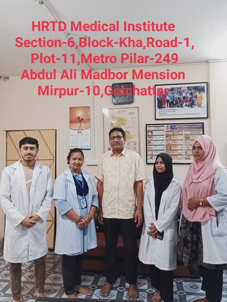
Location of Paramedical course 2 Years.Mobile Number.01987073965.01797522136,HotLine-01969947171 HRTD Medical Institute , Abdul Ali Madbor Mention, Section-6, Block-Kha, Road-1, Plot-11, Mirpur-10 (Gol-Chattar) Metro Rail Pilar NO-249, Dhaka-1216. It is situated by the West Side of Agrani Bank, the South Side of Fire Service, Islami Bank, Janata Bank, Social Islami Bank, Medinova, Ibrahim Diabetic Hospital, the North Side of Baitul Mamur Jame Mosjid, Grave of Baitul Mamur Jame Mosjid, and East Side of Maliha Apartment.
Course Fee For Paramedical Course 2 Years
Admission Fee=16,500/-,Monthly Fee 3000×24=72,000/-,Exam Fee=1000×4=4,000/-, Total Course Fee=92,500/-.Books Fee for Every Semester 1,500/-.
Paramedical Course 2 Years Admission Eligibility
Paramedical Course 2 Years Admission Eligibility. Mobile Number. 01987073965. 01941123488, 01797522136. SSC or Equivalent/HSC/ Degree/ Masters from any Background (Science/ Arts/ Commerce/ Technical).
Documents for Admission in Paramedical Course 2 Years
Paramedical Course 2 Years Course in Dhaka. Mobile No: 01987-073965, 01797-522136. HRTD Medical Institute. Document Needed: Photocopy of Certificate, Photocopy of NID, Passport Size Photo 4 Pcs. Without NID, a Birth Certificate is allowed for an emergency case.
Hostel Facilities in HRTD Medical Institute
Hostel & Meal Facilities
The Institute has hostel facilities for the students. Students can take a bed in the hostel.
Hostel Fee Tk 3000/- Per Month
Meal Charges Tk 3000/- Per Month. ( Approximately )
হোস্টাল ও খাবার সুবিধা
ইনস্টিটিউটে শিক্ষার্থীদের জন্য হোস্টেল সুবিধা রয়েছে। ছাত্ররা হোস্টেলে বিছানা নিতে পারে।
হোস্টেল ফি 3000/- টাকা প্রতি মাসে,
খাবারের চার্জ 3000/- টাকা প্রতি মাসে।(প্রায়)
Teachers For Paramedical Course 2 Years
- Dr. Md. Sakulur Rahman, MBBS, CCD (BIRDEM), Course Director
- Dr. Sanjana Binte Ahmed, BDS, MPH, Assistant Course Director
- Dr. Tisha, MBBS, PGT Gyne, Assistant Course Director
- Dr. Suhana, MBBS, PGT Medicine
- Dr. Danial Hoque, MBBS, C-Card
- Dr. Tisha, MBBS
- Dr. Afrin Jahan, MBBS, PGT Medicine
- Dr. Ananna, MBBS
- Dr. Lamia Afroze, MBBS
- Dr. Amena Afroze Anu, MBBS, PGT Gyne, Assistant Course Director
- Dr. Farhana Antara, MBBS,
- Dr. Nazmun Nahar Juthi, BDS, PGT
- Dr. Farhana Sharna, MBBS
- Dr. Bushra, MBBS
- Dr. Turzo, MBBS
- Dr. Kamrunnahar Keya, BDS, PGT (Dhaka Dental College)
- Dr. Shamima, MBBS, PGT Gyne
- Dr. Alamin, MBBS
- Dr. Benzir Belal, MBBS
- Dr. Disha, MBBS
- Dr. Mahinul Islam, MBBS
- Dr. Tisha, MBBS, PGT Medicine
- Dr. Anika, MBBS, PGT
- Dr. Jannatul Ferdous, MBBS, PGT Gyne
- Dr. Jannatul Aman, MBBS, PGT
- Dr. Rayhan, BPT
- Dr. Abu Hurayra, BPT
- Dr. Sharmin Ankhi, MBBS, PGT Medicine
- Md. Monir Hossain, B Pharm, M Pharm
- Md. Monirul Islam, B Pharm, M Pharm
- Md. Feroj Ahmed, BSc Pathology, PDT Medicine
Practical Class For Paramedical Course 2 Years
- Heart Beat, Heart Rate
- Heart Sound,Pulse
- Blood Pressure, Hypertension, Hypotension
- First Aid Box
- Auscultation
- Inhaler, Rotahaler
- Nebulizer
- Glucometer Blood Glucose
- Injection I/V
- Injection I/M
- Cleaning,Dressing,Bandaging
- Saline
- CPR
- Stitch
- Body Temperature
- Nasal Tube Gel ,Hand Wash
- Blood Grouping
- Cyanosis, Dehydration Test, Edema Test
Subjects for Paramedical Course 2 Years
Paramedical Course 2 Years .This Course Contains 18 Subject in 4 Semester. Mobile Number: 01987073965,01797-522136
1st Semester Subjects
- Human Anatomy & Physiology-1
- Pharmacology-1
- Study of OTC Drugs
- First Aid-1 & 2 & Practice of Medicine
- Hematology & Pathology for Medical Practice
2nd Semester Subjects
- Cardiovascular Anatomy & Physiology
- Medical Biochemistry
- Medical Diagnosis-1
- Surgery & Antimicrobial Drugs
- Orthopedic Anatomy
3rd Semester Subjects
- General Pathology
- Pharmacology-2
- Practice of Medicine-2 & 3
- Essential Drugs
4th Semester Subjects
- Neuro Anatomy & Physiology
- Histology & Cytology
- Respiratory Disease & Treatment
- Practice of Neuro Medicine
Some Practical Class Details Given Below for Paramedical Course 2 Years
Blood Pressure Practical
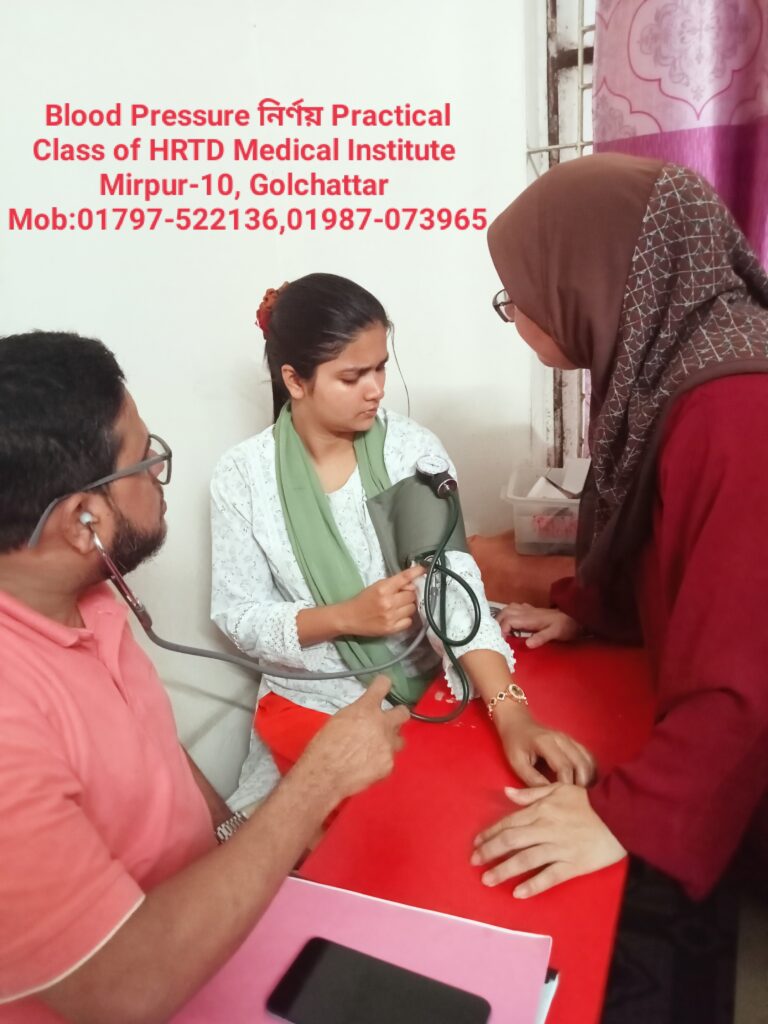
Blood pressure is recorded as two numbers, such as 120/80. The larger number is the pressure in the arteries as the heart pumps out blood during each beat. This is called the systolic blood pressure. The lower number is the pressure as the heart relaxes before the next beat.
Systolic BP / Diastolic BP
Example: 120/80 mmHg
Types of Blood Pressure
1. Systolic Blood Pressure (SBP)
- Pressure during ventricular contraction (systole)
- Normal: 120 mmHg
2. Diastolic Blood Pressure (DBP)
- Pressure during ventricular relaxation (diastole)
- Normal: 80 mmHg
Normal Blood Pressure Values (Adults)
| Category | Systolic (mmHg) | Diastolic (mmHg) |
|---|---|---|
| Normal | 90–120 | 60–80 |
| Pre-Hypertension | 121–139 | 81–89 |
| Hypertension Stage 1 | 140–159 | 90–99 |
| Hypertension Stage 2 | ≥160 | ≥100 |
| Hypotension | <90 | <60 |
Abnormal Blood Pressure
1. Hypertension
Persistently elevated BP
Causes:
- Primary (essential)
- Secondary (renal, endocrine)
Complications:
- Stroke
- Heart attack
- Kidney failure
- Retinopathy
2. Hypotension
Low blood pressure
Causes:
- Dehydration
- Blood loss
- Shock
- Heart failure
Symptoms:
- Dizziness
- Fainting
- Weak pulse
Heart Beat
Definition
A heart beat is one complete cycle of contraction (systole) and relaxation (diastole) of the heart.
- Each beat pumps blood to the body
- Controlled by the cardiac conduction system
Heart Rate (HR)
Definition
Heart Rate is the number of heart beats per minute (bpm).
Example:
- 72 beats/minute = normal adult resting heart rate
Normal Heart Rate Values
Adults
- 60–100 bpm
Children
- 70–120 bpm
Infants
- 120–160 bpm
Athletes
- 40–60 bpm (normal due to strong heart)
Types of Heart Rate
1. Tachycardia
- HR > 100 bpm
- Causes: fever, exercise, stress, anemia
2. Bradycardia
- HR < 60 bpm
- Causes: athletes, hypothermia, heart block
Nebulizer Practical for Paramedical Course 2 Years
Definition
A nebulizer is a medical device that converts liquid medicine into a fine mist (aerosol) so that it can be inhaled directly into the lungs.
Purpose of Nebulizer
- To deliver medicine directly to the respiratory tract
- Useful when patients cannot use inhalers properly
- Provides quick relief in breathing problems
Common Uses
- Asthma
- COPD (Chronic Obstructive Pulmonary Disease)
- Bronchitis
- Pneumonia
- Acute respiratory distress
- Wheezing in children
- Emergency shortness of breath
Types of Nebulizer
1. Jet Nebulizer
- Uses compressed air
- Most commonly used
- Affordable
2. Ultrasonic Nebulizer
- Uses ultrasonic vibrations
- Quiet operation
- Faster drug delivery
3. Mesh Nebulizer
- Uses vibrating mesh
- Portable
- Efficient and expensive
Parts of a Nebulizer
- Air compressor
- Nebulizer cup (medicine chamber)
- Mouthpiece or face mask
- Tubing
Common Drugs Used in Nebulization
- Salbutamol (Albuterol) – bronchodilator
- Ipratropium bromide
- Budesonide
- Normal saline
- Antibiotics (in some cases)
Procedure of Nebulization
- Wash hands
- Measure prescribed medicine
- Pour medicine into nebulizer cup
- Connect tubing to compressor
- Place mask/mouthpiece properly
- Switch on the machine
- Patient breathes slowly and deeply
- Continue until mist stops (5–10 minutes)
- Clean equipment after use
Advantages
- Easy to use
- Suitable for children and elderly
- Direct action on lungs
- Less systemic side effects
Disadvantages
- Bulky (except portable types)
- Requires electricity
- Longer time than inhalers
Side Effects
- Tremors
- Palpitations
- Dry mouth
- Throat irritation
- Rare allergic reactions
Glucometer Blood Glucose Practical For Paramedical Course 2 Years
A glucometer (blood sugar meter) is a portable medical device that measures the amount of sugar (glucose) in a drop of blood, essential for managing diabetes by helping you see how food, exercise, and medicine affect your levels, typically using a finger prick and a test strip to get a reading in mg/dL or mmol/L within seconds. To use, you prick your finger (after washing hands), touch the blood to a test strip, and the meter displays your blood sugar level, guiding treatment and preventing complications.
How it Works
- Prepare: Wash hands, insert a test strip into the meter (this turns it on).
- Lancet: Prepare a lancet device, setting the depth and loading it.
- Prick: Firmly press the lancet on the side of your fingertip and squeeze a small blood drop.
- Apply Blood: Touch the drop to the edge of the test strip.
- Read: The meter counts down and displays the glucose level (e.g., 80-130 mg/dL is a common target before meals).
Key Things to Know
- Purpose: To manage diabetes by tracking glucose levels, preventing highs (hyperglycemia) and lows (hypoglycemia).
- Components: A handheld meter, disposable test strips, and lancets.
- Accuracy: Store strips properly (sealed, room temp) and check expiration dates; a small drop of blood is needed.
- When to Use: People with diabetes use them multiple times daily to guide treatment.
Target Ranges (General Guidelines)
- Before Meals (Preprandial): 80-130 mg/dL.
- After Meals (Postprandial): Less than 180 mg/dL (2 hours after starting to eat).
By monitoring these numbers, you and your healthcare provider can adjust diet, exercise, and medication to keep your blood sugar in a healthy range.
Some Subject Details for Paramedical Course 2 Years
Human Anatomy & Physiology for Paramedical Course 2 Years
The Study of the body Structure and its function is Anatomy & Physiology. Here we discuss the systems of the human body and its organ, Tissues, and cells. The systems of the human body are the digestive system, Respiratory system, Cardiovascular system, Skeletal system, Muscular system, nervous system, Endocrine system, Immune System, Integumentary System and Urinary System.
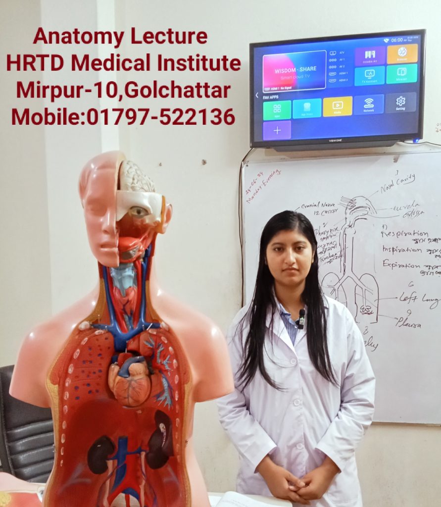
Anatomy is the study of body structures and their relationships, while physiology is the study of how those structures function; they are interconnected, as form dictates function (e.g., the structure of a neuron’s axon allows it to transmit signals). Together, they explain the human body’s workings, from cells and tissues to organ systems, maintaining balance (homeostasis) through complex processes like temperature regulation, making them fundamental to medicine and health sciences.
Anatomy (The “What” & “Where”)
- Definition: The study of the body’s physical structures, both internal and external.
- Levels: Explores structures from microscopic (cells, tissues) to macroscopic (organs, organ systems).
- Types: Can be gross (large structures), microscopic (histology/cytology), regional (by body area), or systemic (by body system).
Physiology (The “How”)
- Definition: The study of the functions and processes of body parts and systems.
- Focus: Explains how structures work together to keep the body alive, such as the circulatory system delivering oxygen or the integumentary system protecting the body.
The Interconnection: Form Meets Function
- The principle “form fits function” is key: a structure’s anatomy enables its physiological role.
- Example: Dendrites (anatomy) receive signals, allowing the neuron (physiology) to function as a receiver, while the axon (anatomy) transmits signals (physiology).
Key Concepts in A&P
- Homeostasis: The body’s ability to maintain a stable internal environment (e.g., regulating temperature).
- Body Systems: The interconnected network of organs (e.g., cardiovascular, nervous, integumentary) working cooperatively.
- Anatomical Position: A standard reference posture (standing upright, palms forward) used to describe body locations universally.
Major Systems of the Human Body
| System | Main Function |
|---|---|
| Skeletal | Support & protection |
| Muscular | Movement |
| Nervous | Control & coordination |
| Endocrine | Hormonal regulation |
| Cardiovascular | Blood circulation |
| Respiratory | Gas exchange |
| Digestive | Nutrition & digestion |
| Urinary | Waste excretion |
| Reproductive | Reproduction |
| Integumentary | Protection & temperature control |
Relationship Between Anatomy & Physiology
| Anatomy | Physiology |
|---|---|
| Structure | Function |
| Form | Action |
| Static study | Dynamic study |
কয়েকটি সিস্টেম সম্পর্কে আলোচনা করা হলো:
1. Skeletal System
Function:
- Gives shape and support to the body
- Protects vital organs (brain, heart, lungs)
- Helps in body movement
- Produces blood cells in bone marrow
Main Parts:
- Bones (206 bones)
- Joints
- Cartilage
2. Muscular System
Function:
- Enables movement of the body
- Maintains posture
- Produces heat
Types of Muscles:
- Skeletal muscle – voluntary
- Smooth muscle – involuntary
- Cardiac muscle – found in the heart
3. Nervous System
Function:
- Controls and coordinates body activities
- Receives and responds to stimuli
- Responsible for thinking, memory, and emotions
Main Parts:
- Brain
- Spinal cord
- Nerves
4. Cardiovascular System
Function:
- Circulates blood throughout the body
- Transports oxygen, nutrients, and hormones
- Removes waste products
Main Parts:
- Heart
- Blood
- Blood vessels (arteries, veins, capillaries)
5. Respiratory System
Function:
- Helps in breathing
- Supplies oxygen to the blood
- Removes carbon dioxide from the body
Main Parts:
- Nose
- Trachea
- Lungs
- Alveoli
6. Digestive System
Function:
- Digestion of food
- Absorption of nutrients
- Elimination of waste
Main Parts:
- Mouth
- Esophagus
- Stomach
- Intestines
- Liver and pancreas
Pharmacology-1 for Paramedical Course 2 Years
1. Definition of Pharmacology
Pharmacology is the branch of medical science that deals with drugs, their sources, actions, uses, side effects, and mechanisms in the human body.
2. Drug – Definition
A drug is a chemical substance that is used to diagnose, prevent, treat, or cure disease.
3. Branches of Pharmacology
- Pharmacokinetics – What the body does to the drug
- Pharmacodynamics – What the drug does to the body
- Pharmacotherapeutics – Use of drugs in treatment
- Toxicology – Study of harmful effects of drugs
- Chemotherapy – Drugs used to treat infections and cancer
4. Pharmacokinetics (ADME)
A – Absorption:
How a drug enters the bloodstream
D – Distribution:
How the drug spreads in the body
M – Metabolism:
Breakdown of drugs mainly in the liver
E – Excretion:
Removal of drugs mainly through kidneys (urine)
5. Pharmacodynamics
- Drug action and effect
- Receptor interaction
- Dose–response relationship
Example:
Paracetamol reduces pain and fever.
6. Routes of Drug Administration
- Oral (by mouth)
- Sublingual
- Intravenous (IV)
- Intramuscular (IM)
- Subcutaneous (SC)
- Topical
- Inhalation
7. Types of Drugs
- Analgesics (pain killers)
- Antibiotics
- Antipyretics (reduce fever)
- Antiseptics
- Sedatives
8. Adverse Drug Reactions (ADR)
- Nausea
- Vomiting
- Allergy
- Drowsiness
9. Importance of Pharmacology for Nurses / Physiotherapists
- Safe drug administration
- Understanding drug effects
- Prevention of medication errors
- Patient education
10. Common Terms
- Dose: Amount of drug given
- Therapeutic effect: Desired effect
- Side effect: Unwanted effect
- Overdose: Excess amount of drug
Study of OTC Drugs for Paramedical Course 2 Years
OTC Drugs are important for all Medical Assistant Courses. These Drugs are Emergency and safe for the patients. The Study of OTC Drugs improves the quality of practice. Some OTC Drugs are Albendazole, Ascorbic Acid, Calcium, Multivitamins, Vitamin B Complex, Omeprazole, Oral Rehydration Salt, Salbutamol etc.
Definition
OTC drugs are medicines that can be purchased without a doctor’s prescription and are used for minor and self-limiting illnesses.
Purpose of OTC Drugs
- Relief of common symptoms
- Easy access to treatment
- Cost-effective healthcare
- Reduces burden on hospitals
Characteristics of OTC Drugs
- Safe when used as directed
- Low risk of serious side effects
- Clear labeling and instructions
- Suitable for self-medication
Classification of OTC Drugs
1. Analgesics & Antipyretics
Used for pain and fever.
| Drug | Use |
|---|---|
| Paracetamol | Fever, mild pain |
| Ibuprofen | Pain, inflammation |
2. Antacids
Used for acidity and indigestion.
| Drug | Use |
|---|---|
| Aluminum hydroxide | Neutralizes acid |
| Magnesium hydroxide | Relieves acidity |
3. Cough & Cold Preparations
- Antitussives – suppress cough
- Expectorants – remove mucus
- Decongestants – relieve nasal block
Examples:
- Dextromethorphan
- Guaifenesin
- Phenylephrine
4. Antihistamines
Used for allergy.
| Drug | Use |
|---|---|
| Cetirizine | Allergic rhinitis |
| Loratadine | Urticaria |
5. Antidiarrheal Drugs
Used for diarrhea.
| Drug | Use |
|---|---|
| ORS | Prevents dehydration |
| Loperamide | Reduces bowel movement |
6. Laxatives
Used for constipation.
- Bulk-forming: Isabgol
- Osmotic: Lactulose
- Stool softeners
7. Topical Preparations
Used on skin.
- Antiseptics (Povidone-iodine)
- Antifungal creams
- Pain relief gels
8. Vitamins & Minerals
- Vitamin B-complex
- Vitamin C
- Iron supplements
Dosage Forms of OTC Drugs
- Tablets
- Syrups
- Capsules
- Creams & ointments
- Drops
- Powders
Advantages of OTC Drugs
- Easily available
- Saves time & money
- Suitable for minor illnesses
- Promotes self-care
First Aid Paramedical Course 2 Years
First Aid is an important subject for Medical courses including Paramedical. RMP Courses, LMAF Courses, Paramedical Courses, DMA Courses, DMS Courses, Nursing Courses, Dental Courses, Pathology Courses, Physiotherapy Courses, Caregiver courses etc. Here we discuss shock, Classification Shock, causes of Shock, Stages of Shock, Clinical Features of Shock, Hypovolemic Shock, Cardiogenic Shock, Neurogenic Shock, Traumatic Shock, Burn Shock, Electric Shock, Psychogenic Shock, Anaphylactic Shock, First Aid of Shock, First Aid of cut, First Aid of Snake Bite, First Aid of Accidental Injury etc.
Definition
First Aid is the immediate and temporary care given to an injured or suddenly ill person before professional medical treatment is available.
Aims of First Aid
- Preserve life
- Prevent condition from worsening
- Promote recovery
Principles of First Aid
- Stay calm
- Ensure safety of the scene
- Assess the casualty
- Call for medical help
- Provide appropriate care
- Do not give unnecessary treatment
First Aid – Primary Survey (DRABC)
| Step | Meaning |
|---|---|
| D | Danger – ensure safety |
| R | Response – check consciousness |
| A | Airway – clear airway |
| B | Breathing – check breathing |
| C | Circulation – control bleeding |
Basic First Aid Procedures
1. Wounds & Bleeding
- Apply direct pressure
- Elevate the injured part
- Use clean dressing
- Do not remove embedded objects
2. Burns
- Cool with running water (10–20 minutes)
- Cover with sterile dressing
- Do not apply oil or ointment
- Do not burst blisters
3. Fractures
- Immobilize the injured part
- Use splints
- Do not move unnecessarily
- Seek medical help
4. Fainting
- Lay patient flat
- Raise legs
- Loosen tight clothing
- Ensure fresh air
5. Nose Bleeding (Epistaxis)
- Sit patient upright
- Lean forward
- Pinch nostrils for 10 minutes
- Do not tilt head backward
6. Choking
- Encourage coughing
- Give back blows
- Perform abdominal thrusts (Heimlich maneuver)
7. Poisoning
- Identify poison if possible
- Do not induce vomiting (unless instructed)
- Seek emergency care immediately
8. Snake Bite
- Keep patient calm
- Immobilize limb
- Do not cut or suck wound
- Take patient to hospital urgently
CPR (Cardiopulmonary Resuscitation)
Steps
- Check responsiveness
- Call emergency help
- Start chest compressions
- Give rescue breaths
Compression : Breath ratio = 30 : 2
First Aid Kit – Contents
- Sterile gauze
- Bandages
- Antiseptic solution
- Adhesive tape
- Scissors
- Gloves
- Pain relievers
- ORS packets
Practice of Medicine for Paramedical Course 2 Years
Diploma In Paramedical Course 2 Years for Important topics in the practice of medicine include fundamental sciences like anatomy, physiology, and pathology; core clinical subjects such as internal medicine, surgery, and pediatrics; and practical applications like pharmacology, diagnostic procedures, and patient management. Additionally, crucial areas include public health (sanitation, vaccination), family medicine (addressing a wide range of patient needs), and increasingly, community medicine (epidemiology, health indicators), and interdisciplinary fields like addiction medicine, forensic medicine, and genomic medicine.
Definition
Practice of Medicine is the branch of medical science concerned with the prevention, diagnosis, treatment, and management of diseases in adult patients through clinical knowledge and patient care.
Objectives of Practice of Medicine
- Diagnose diseases accurately
- Treat and manage medical conditions
- Prevent complications
- Promote health and recovery
- Provide holistic patient care
Scope of Practice of Medicine
Includes:
- History taking
- Physical examination
- Clinical diagnosis
- Investigations
- Medical management
- Follow-up care
Components of Practice of Medicine
1. History Taking
Includes:
- Chief complaints
- History of present illness
- Past medical history
- Drug history
- Family history
- Social history
2. Physical Examination
- General examination
- Systemic examination:
- Cardiovascular system
- Respiratory system
- Gastrointestinal system
- Nervous system
3. Diagnosis
- Provisional diagnosis
- Differential diagnosis
- Final diagnosis
4. Investigations
- Blood tests
- Urine tests
- X-ray
- ECG
- Ultrasound
- CT/MRI (when needed)
5. Treatment
- Drug therapy
- Dietary advice
- Lifestyle modification
- Supportive care
Common Diseases Studied in Practice of Medicine
Cardiovascular Diseases
- Hypertension
- Ischemic heart disease
- Heart failure
Respiratory Diseases
- Asthma
- COPD
- Pneumonia
- Tuberculosis
Gastrointestinal Diseases
- Peptic ulcer
- Hepatitis
- Diarrhea
Endocrine Diseases
- Diabetes mellitus
- Thyroid disorders
Neurological Diseases
- Stroke
- Epilepsy
- Parkinson’s disease
Infectious Diseases
- Malaria
- Dengue
- Typhoid
- COVID-19
Hematology for Paramedical Course 2 Years
Hematology is the branch of medicine focused on the study, diagnosis, and treatment of blood, blood-forming organs (like bone marrow, spleen, lymph nodes), and blood disorders, including anemias, clotting issues (hemophilia), and blood cancers (leukemia, lymphoma).
What hematology covers:
- Blood Components: Red cells (oxygen), white cells (immunity), platelets (clotting), plasma, and proteins.
- Blood-Forming Organs: Bone marrow, lymph nodes, spleen, thymus.
- Disorders: Anemia, bleeding disorders (hemophilia), clotting disorders, blood cancers, and immune issues.
- Processes: Blood formation (hematopoiesis) and coagulation (clotting).
Who are the specialists?
- Hematologist: A physician (often an internist or pediatrician) who diagnoses and treats patients with blood disorders, managing their care directly.
- Hematopathologist: A pathologist who specializes in analyzing blood, bone marrow, and lymphoid tissues in a lab to diagnose diseases.
- Hematology-Oncology: Many hematologists train in both areas, as blood cancers are a major focus.
Common conditions treated:
- Anemia (e.g., sickle cell anemia)
- Bleeding & Clotting Disorders (e.g., Hemophilia, Thrombosis)
- Blood Cancers (e.g., Leukemia, Multiple Myeloma, Lymphoma)
- Immune deficiencies related to blood.
Composition of Blood
Total blood volume ≈ 5–6 liters in adults.
A. Plasma (≈55%)
Plasma is the liquid part of blood.
Components:
- Water (90–92%)
- Plasma proteins
- Albumin – maintains osmotic pressure
- Globulin – immunity (antibodies)
- Fibrinogen – blood clotting
- Electrolytes – Na⁺, K⁺, Ca²⁺
- Nutrients – glucose, amino acids, lipids
- Hormones & enzymes
- Waste products – urea, creatinine
Functions of Plasma:
- Maintains blood pressure
- Transport medium
- Helps in clotting and immunity
Formed Elements of Blood
A. Red Blood Cells (RBCs / Erythrocytes)
Structure:
- Biconcave, non-nucleated
- Diameter ≈ 7.5 µm
Normal Count:
- Male: 5–6 million/mm³
- Female: 4–5 million/mm³
Hemoglobin (Hb):
- Iron-containing protein
- Carries oxygen
Life Span:
- Approximately 120 days
Functions:
- Oxygen transport
- Carbon dioxide transport
- Acid–base balance
RBC Disorders:
- Anemia – low RBC or Hb
- Polycythemia – increased RBC count
B. White Blood Cells (WBCs / Leukocytes)
Normal Count:
- 4,000–11,000/mm³
Function:
- Body defense and immunity
Types of WBCs
1. Neutrophils (60–70%)
- First line of defense
- Fight bacterial infection
2. Lymphocytes (20–30%)
- B cells – antibody production
- T cells – cell-mediated immunity
3. Monocytes (2–8%)
- Phagocytosis
- Become macrophages
4. Eosinophils (1–4%)
- Allergic reactions
- Parasitic infections
5. Basophils (0.5–1%)
- Release histamine
- Inflammation and allergy
WBC Disorders:
- Leukocytosis – increased WBC
- Leukopenia – decreased WBC
- Leukemia – cancer of blood cells
C. Platelets (Thrombocytes)
Normal Count:
- 150,000–400,000/mm³
Life Span:
- 7–10 days
Function:
- Blood clotting
- Prevention of bleeding
Disorder:
- Thrombocytopenia – low platelet count
Hemostasis (Blood Clotting)
Steps:
- Vasoconstriction
- Platelet plug formation
- Coagulation
- Fibrinogen → Fibrin (clot formation)
Importance:
- Prevents excessive blood loss
Hemoglobin (Hb) in Detail
Normal Values:
- Male: 13–18 g/dL
- Female: 12–16 g/dL
Functions:
- Oxygen transport
- Maintains blood pH
Abnormalities:
- Low Hb → anemia
- High Hb → polycythemia
Blood Groups
ABO Blood Group System
- Group A
- Group B
- Group AB
- Group O
Rh Factor
- Rh positive
- Rh negative
Importance:
- Safe blood transfusion
- Pregnancy (Rh incompatibility)
Blood Formation (Hematopoiesis)
Site:
- Bone marrow (main site)
Process:
- Stem cells → RBCs, WBCs, platelets
Clinical Importance of Hematology
- Diagnosis of anemia and infections
- Monitoring treatment response
- Blood transfusion safety
- Management of bleeding disorders
Pathology for Paramedical Course 2 Years
Pathology is the medical science of studying diseases—their causes, mechanisms, and effects—primarily by examining tissues, cells, and body fluids (like blood/urine) in labs to diagnose illnesses, guide treatment, and monitor health, acting as the crucial link between basic science and clinical medicine, often involving microscopic analysis of biopsies, genetic testing, and microbiology. Pathologists are doctors specializing in this lab-based diagnosis, making critical decisions for cancer, infections, and chronic diseases, even performing autopsies to understand death.
Key Aspects of Pathology
- Study of Disease: Investigates how diseases start (etiology) and develop (pathogenesis).
- Diagnostic Focus: Analyzes samples (biopsies, blood, urine) to find abnormalities.
- Core Disciplines: Includes histology (tissues), cytology (cells), microbiology (germs), clinical chemistry, and molecular pathology (genetics).
- Tools & Techniques: Uses microscopes, special stains, immunological markers, and genetic tests.
- Clinical Role: Provides vital information for surgeons, oncologists, and other clinicians to diagnose, treat, and manage conditions like cancer, infections, and autoimmune disorders.
What Pathologists Do
- Examine Specimens: Look at tissue under a microscope for signs of cancer, inflammation, or infection.
- Perform Tests: Analyze blood for chemical imbalances or infectious agents.
- Consult: Work with other doctors to determine the best course of treatment.
- Conduct Autopsies: Investigate deaths to determine cause and manner.
Modern Advancements
- Digital Pathology: Digitizing slides for easier sharing and analysis.
- AI in Diagnostics: Using artificial intelligence to spot patterns and improve accuracy.
- Molecular Pathology: Analyzing DNA and RNA for targeted therapies, especially in cancer.
1. Definition
Pathology is the branch of medical science that studies:
- The causes of disease (etiology)
- The mechanism of disease development (pathogenesis)
- The structural and functional changes in tissues and organs (morphology)
- The effects of disease on the body
বাংলা সংজ্ঞা:
প্যাথলজি হলো রোগের কারণ, বিকাশ প্রক্রিয়া, অঙ্গ–প্রত্যঙ্গে পরিবর্তন এবং দেহে তার প্রভাবের অধ্যয়ন।
2. Branches of Pathology
- General Pathology – Study of disease processes common to all organs (e.g., inflammation, necrosis)
- Systemic Pathology – Study of diseases of specific organs or systems (e.g., cardiovascular, respiratory)
- Clinical Pathology – Laboratory study of blood, urine, body fluids for diagnosis
- Forensic Pathology – Study of death, injury, and crime-related pathology
- Molecular Pathology – Study of diseases at cellular and molecular level
3. Etiology (Causes of Disease)
- Genetic – inherited conditions (e.g., sickle cell anemia)
- Infectious – bacteria, viruses, fungi, parasites
- Physical – trauma, burns, radiation
- Chemical – poisons, toxins, drugs
- Nutritional – deficiency or excess (e.g., scurvy, obesity)
- Immune – autoimmune diseases
4. Pathogenesis (Mechanism of Disease)
- How the disease develops in the body
- Example: Atherosclerosis
- Fat deposits in arteries → narrowing → reduced blood flow → heart attack
5. Morphological Changes
- Gross Changes: visible changes in organs (e.g., enlarged liver)
- Microscopic Changes: cellular changes under microscope (e.g., inflammation, necrosis)
6. Common Pathological Processes
- Inflammation – Body’s response to injury or infection
- Degeneration – Deterioration of cells
- Necrosis – Cell death
- Neoplasia – Uncontrolled cell growth (benign or malignant)
- Hemodynamic Disorders – Bleeding, clotting, edema
- Infections – Bacterial, viral, fungal, parasitic.
Cardiovascular Anatomy for Paramedical Course 2 Years
Cardiovascular Nursing involves the heart (a four-chambered pump), blood vessels (arteries, veins, capillaries), and blood, forming circuits to deliver oxygen/nutrients and remove waste, divided into pulmonary (heart-lungs) and systemic (heart-body) loops, crucial for body function. Key structures include the atria (receiving chambers), ventricles (pumping chambers), four valves (tricuspid, pulmonary, mitral, aortic) controlling flow, and major vessels like the aorta, vena cavae, and pulmonary arteries/veins, all working to circulate life-sustaining blood.
The Heart: The Central Pump
- Chambers: Four chambers: Right Atrium, Right Ventricle, Left Atrium, Left Ventricle.
- Valves: Four one-way valves ensure proper blood flow: Tricuspid, Pulmonary, Mitral (Bicuspid), Aortic.
- Septum: Muscular walls separating the right and left sides (interatrial and interventricular septum).
Blood Vessels: The Network
- Arteries: Carry oxygenated blood away from the heart (e.g., Aorta, Pulmonary Artery).
- Veins: Return deoxygenated blood to the heart (e.g., Vena Cavae, Pulmonary Veins).
- Capillaries: Tiny vessels connecting arterioles and venules, where nutrient/gas exchange occurs.
The Two Circuits
- Pulmonary Circulation: Right side of the heart pumps deoxygenated blood to the lungs to get oxygen, then back to the left heart.
- Systemic Circulation: Left side pumps oxygenated blood to the body’s tissues, and deoxygenated blood returns to the right heart.
Blood Flow Through the Heart (Simplified)
- Deoxygenated blood enters Right Atrium (from Vena Cavae).
- Right Atrium to Right Ventricle (through Tricuspid Valve).
- Right Ventricle to Lungs (through Pulmonary Valve & Artery).
- Oxygenated blood returns to Left Atrium (from Pulmonary Veins).
- Left Atrium to Left Ventricle (through Mitral Valve).
- Left Ventricle pumps to the body (through Aortic Valve & Aorta).
Orthopedic Anatomy for Paramedical Course 2 Years
Orthopedic anatomy is the study of the musculoskeletal system, the complex network of tissues that provides the body with structure, support, and the ability to move. This field forms the foundation of orthopedic surgery and physical therapy, focusing on how these components interact to withstand stress and perform mechanical work.
Core Components of the Musculoskeletal System
The orthopedic system is comprised of several distinct types of tissue, each with specific structural roles:
- Bones: The adult human skeleton consists of 206 bones. They provide the rigid framework for the body and protect internal organs.
- Joints: There are approximately 230 joints in the adult body where two or more bones meet. They are classified by their movement type, such as hinge (elbow), ball-and-socket (hip/shoulder), or saddle (thumb).
- Muscles: The body contains roughly 640 skeletal muscles. These voluntary muscles contract to pull on bones, facilitating movement.
- Ligaments: Dense bands of fibrous tissue that connect bone to bone, providing stability to joints.
- Tendons: Fibrous cords that connect muscle to bone, transmitting the force of muscle contraction to move the skeleton.
- Cartilage: A smooth, resilient tissue that covers the ends of bones at joints (articular cartilage) to reduce friction.
Anatomical Regions in Orthopedics
Orthopedists often specialize in specific regions due to their complex anatomical requirements:
- Upper Limb: Includes the shoulder, arm (humerus), forearm (radius/ulna), and the intricate bones of the hand and wrist.
- Lower Limb: Encompasses the hip, thigh (femur), knee, leg (tibia/fibula), and the foot and ankle.
- Spine: Focuses on the vertebrae, intervertebral discs, and the spinal cord, which are critical for axial stability and neurological health.
Clinical Significance
Understanding these structures is essential for diagnosing and treating common orthopedic conditions:
- Fractures: Breaks in the bone, often described by their location (e.g., distal, proximal) and displacement.
- Sprains vs. Strains: A sprain refers to an injury of a ligament (bone-to-bone), while a strain refers to an injury of a muscle or tendon (muscle-to-bone).
- Arthritis: The degradation of articular cartilage, leading to pain and reduced joint mobility.
