
Diploma Physiotherapy Assistant(DPTA) Course 2 Years Details
Diploma Physiotherapy Assistant Course 2 Years. Details: Mobile Number-01987073965,01797522136.This Course Contains 18 Subject. There are Total 4 Semester. The 1st Semester Contains 5 Subject Which are Human Anatomy & Physiology, Chemistry & Pharmacology, Study of OTC, First Aid & Practice Of Medicine, Hematology & Pathology. 2nd Semester Contains 5 Subject Which are Cardiovascular Anatomy, Orthopedic Anatomy ,Neuro Anatomy, Biochemistry, Electro Physics.3rd Semester Contains 4 Subject And The Last Semester also Contains 4 Subject.
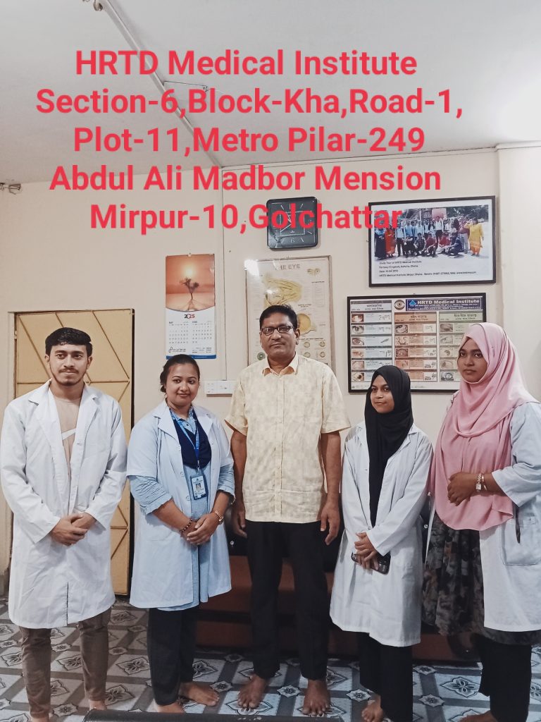
Location of Diploma Physiotherapy Assistant Course 2 Years
Diploma Physiotherapy Assistant Course 2 Years. Mobile Number.01987073965,01797522136. HRTD Medical Institute , Abdul Ali Madbor Mention, Section-6, Block-Kha, Road-1, Plot-11, Mirpur-10 (Gol-Chattar) Metro Rail Pilar NO-249, Dhaka-1216. It is situated by the West Side of Agrani Bank, the South Side of Fire Service, Islami Bank, Janata Bank, Social Islami Bank, Medinova, Ibrahim Diabetic Hospital, the North Side of Baitul Mamur Jame Mosjid, Grave of Baitul Mamur Jame Mosjid, and East Side of Maliha Apartment.
Orthopedic Lecture Dr.Anu
Course Fee For Diploma Physiotherapy Assistant Course 2 Years
DPTA Course 2 Years Admission Fee: 16,500/-,Monthly Fee 3000×24=72,000/-,Exam Fee=1000×4=4,000/-, Total Course Fee=92,500/-.Books Fee for Every Semester 1,500/-.
Diploma Physiotherapy Assistant Course 2 Years Admission Eligibility
Diploma Physiotherapy Assistant Course 2 Years Admission Eligibility. Mobile Number. 01987073965. 01941123488, 01797522136. SSC or Equivalent/HSC/ Degree/ Masters from any Background (Science/ Arts/ Commerce/ Technical).
Documents for Admission in Diploma In Physiotherapy Assistant Course 2 Years
Diploma In Physiotherapy Assistant Course 2 Years Course in Dhaka. Mobile No: 01987-073965, 01797-522136. HRTD Medical Institute. Document Needed: Photocopy of Certificate, Photocopy of NID, Passport Size Photo 4 Pcs. Without NID, a Birth Certificate is allowed for an emergency case.
Hostel Facilities in HRTD Medical Institute
Hostal & Meal Facilities
The Institute has hostel facilities for the students. Students can take a bed in the hostel.
Hostel Fee Tk 3000/- Per Month
Meal Charges Tk 3000/- Per Month. ( Approximately )
হোস্টাল ও খাবার সুবিধা
ইনস্টিটিউটে শিক্ষার্থীদের জন্য হোস্টেল সুবিধা রয়েছে। ছাত্ররা হোস্টেলে বিছানা নিতে পারে।
হোস্টেল ফি 3000/- টাকা প্রতি মাসে,
খাবারের চার্জ 3000/- টাকা প্রতি মাসে।(প্রায়)
All Physiotherapy Courses in Dhaka of HRTD Medical Institute
Other Physiotherapy Courses Except for the Diploma Physiotherapy Assistant Course 2 Years. We have others Physiotherapy Courses of Duration 2 Years, 3 Years, and 4 Years. These courses are more valuable and demandable not only in Bangladesh but also over the world. Physiotherapy Course 1 Year Tk 62500/-, Physiotherapy Course 2 Years Tk 92500/-, Physiotherapy Course 3 Years Tk 152500/-, and Physiotherapy Course 4 Years Tk 198500/-. Payment System: Admission Fee, Monthly Fee, and Exam Fee.
Teachers For Diploma Physiotherapy Assistant Course 2 Years
- Dr. Rayhan, BPT
- Dr. Md. Sakulur Rahman, MBBS, CCD (BIRDEM), Course Director
- Dr. Abu Hurayra, BPT
- Dr. Sanjana Binte Ahmed, BDS, MPH, Assistant Course Director
- Dr. Tisha, MBBS, PGT Gyne, Assistant Course Director
- Dr. Suhana, MBBS, PGT Medicine
- Dr. Danial Hoque, MBBS, C-Card
- Dr. Tisha, MBBS
- Dr. Afrin Jahan, MBBS, PGT Medicine
- Dr. Ananna, MBBS
- Dr. Lamia Afroze, MBBS
- Dr. Amena Afroze Anu, MBBS, PGT Gyne, Assistant Course Director
- Dr. Farhana Antara, MBBS,
- Dr. Nazmun Nahar Juthi, BDS, PGT
- Dr. Farhana Sharna, MBBS
- Dr. Bushra, MBBS
- Dr. Turzo, MBBS
- Dr. Kamrunnahar Keya, BDS, PGT (Dhaka Dental College)
- Dr. Shamima, MBBS, PGT Gyne
- Dr. Alamin, MBBS
- Dr. Benzir Belal, MBBS
- Dr. Disha, MBBS
- Dr. Mahinul Islam, MBBS
- Dr. Tisha, MBBS, PGT Medicine
- Dr. Anika, MBBS, PGT
- Dr. Jannatul Ferdous, MBBS, PGT Gyne
- Dr. Jannatul Aman, MBBS, PGT
- Dr. Sharmin Ankhi, MBBS, PGT Medicine
- Md. Monir Hossain, B Pharm, M Pharm
- Md. Monirul Islam, B Pharm, M Pharm
- Md. Feroj Ahmed, BSc Pathology, PDT Medicine
Subjects For Diploma Physiotherapy Assistant Course 2 Years
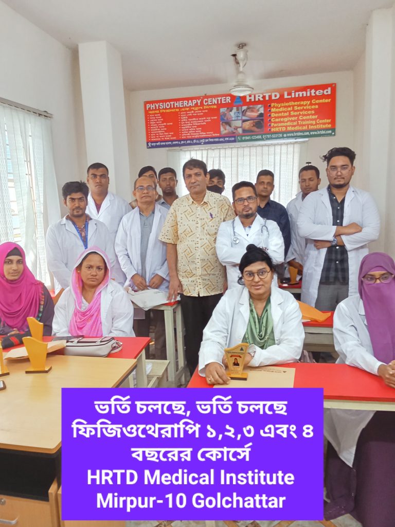
Diploma Physiotherapy Assistant Course 2 Years Contains 18 Subjects. In 2 Years Course There are 4 Semester. Each Semester Duration 6 Months. Total Mobile Number-01987073965,01797522136. HRTD Medical Institute, Mirpur-10, Golchattar.
1st Semester Subjects
- Human Anatomy & Physiology
- Chemistry & Pharma-1
- Study of OTC
- First Aid & Practice of Medicine
- Hematology & Pathology
2nd Semester Subjects
- Cardiovascular Anatomy
- Orthopedic Anatomy
- Neuro Anatomy
- Electro Physics
- Electro Therapy
3rd Semester Subjects
- Human Microbiology
- Bones Joints & Treatment
- Claytons Electrotherapy
- Therapeutic Exercise
4th Semester Subjects
- Orthopedic Disease & Treatment
- Neuropathic Pain & Management
- Electrotherapy-2
- Therapeutic Exercise-2
Practical Classes For Diploma Physiotherapy Assistant Course 2 Years
1st Semester Practical Class For Diploma Physiotherapy Assistant Course 2 Years
- Heart Beat, Heart Rate
- Heart Sound,Pulse
- Blood Pressure, Hypertension, Hypotension
- First Aid Box
- Auscultation
- Inhaler, Rotahaler
- Nebulizer
- Glucometer Blood Glucose
- Injection I/V
- Injection I/M
- Cleaning,Dressing,Bandaging
- Saline
- CPR
- Stitch
- Body Temperature
- Nasal Tube Gel ,Hand Wash
- Blood Grouping
- Cyanosis, Dehydration Test, Edema Test
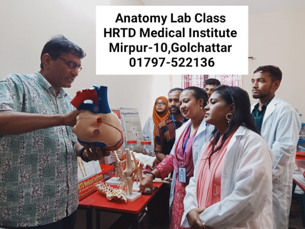
Some Subject Details For Diploma Physiotherapy Assistant 2 Years
Human Anatomy & Physiology Diploma Physiotherapy Assistant 2 Years
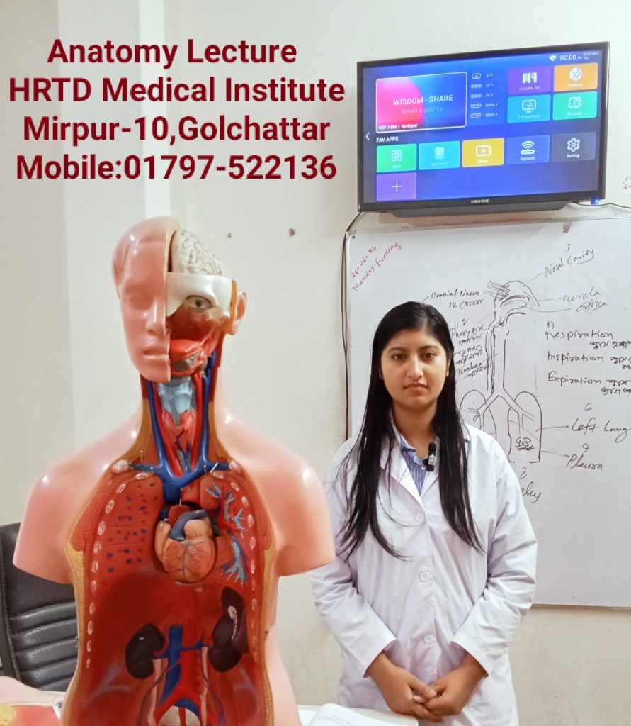
Anatomy is the study of body structures and their relationships, while physiology is the study of how those structures function; they are interconnected, as form dictates function (e.g., the structure of a neuron’s axon allows it to transmit signals). Together, they explain the human body’s workings, from cells and tissues to organ systems, maintaining balance (homeostasis) through complex processes like temperature regulation, making them fundamental to medicine and health sciences.
Definition
Anatomy (English):
Anatomy is the study of the structure of the human body and the relationship between body parts.
অ্যানাটমি (বাংলা):
অ্যানাটমি হলো মানবদেহের গঠন, অঙ্গ-প্রত্যঙ্গ এবং তাদের পারস্পরিক সম্পর্কের অধ্যয়ন।
Physiology (English):
Physiology is the study of the functions of the human body and how body parts work.
ফিজিওলজি (বাংলা):
ফিজিওলজি হলো মানবদেহের বিভিন্ন অঙ্গ কীভাবে কাজ করে তার অধ্যয়ন।
Levels of Organization (শরীরের গঠনের স্তর)
- Cell (কোষ) – দেহের সবচেয়ে ছোট জীবিত একক
- Tissue (টিস্যু) – একই ধরনের কোষের সমষ্টি
- Organ (অঙ্গ) – বিভিন্ন টিস্যু দিয়ে গঠিত (যেমন: হৃদপিণ্ড)
- System (সিস্টেম) – একাধিক অঙ্গের সমন্বয়
- Organism (সম্পূর্ণ মানবদেহ)
Major Systems of Human Body
| System | Main Function |
|---|---|
| Skeletal System | দেহকে আকৃতি ও সাপোর্ট দেয় |
| Muscular System | চলাচল ও শক্তি উৎপাদন |
| Nervous System | দেহের নিয়ন্ত্রণ ও সমন্বয় |
| Cardiovascular System | রক্ত পরিবহন |
| Respiratory System | শ্বাস-প্রশ্বাস |
| Digestive System | খাদ্য হজম |
| Urinary System | বর্জ্য নির্গমন |
| Endocrine System | হরমোন নিঃসরণ |
| Reproductive System | প্রজনন |
| Integumentary System | ত্বক ও সুরক্ষা |
কয়েকটি সিস্টেম সম্পর্কে আলোচনা করা হলো:
1. Skeletal System
Function:
- Gives shape and support to the body
- Protects vital organs (brain, heart, lungs)
- Helps in body movement
- Produces blood cells in bone marrow
Main Parts:
- Bones (206 bones)
- Joints
- Cartilage
2. Muscular System
Function:
- Enables movement of the body
- Maintains posture
- Produces heat
Types of Muscles:
- Skeletal muscle – voluntary
- Smooth muscle – involuntary
- Cardiac muscle – found in the heart
3. Nervous System
Function:
- Controls and coordinates body activities
- Receives and responds to stimuli
- Responsible for thinking, memory, and emotions
Main Parts:
- Brain
- Spinal cord
- Nerves
4. Cardiovascular System
Function:
- Circulates blood throughout the body
- Transports oxygen, nutrients, and hormones
- Removes waste products
Main Parts:
- Heart
- Blood
- Blood vessels (arteries, veins, capillaries)
5. Respiratory System
Function:
- Helps in breathing
- Supplies oxygen to the blood
- Removes carbon dioxide from the body
Main Parts:
- Nose
- Trachea
- Lungs
- Alveoli
6. Digestive System
Function:
- Digestion of food
- Absorption of nutrients
- Elimination of waste
Main Parts:
- Mouth
- Esophagus
- Stomach
- Intestines
- Liver and pancreas
Pharmacology-1 Diploma Physiotherapy Assistant 2 Years
1. Definition of Pharmacology
Pharmacology is the branch of medical science that deals with drugs, their sources, actions, uses, side effects, and mechanisms in the human body.
2. Drug – Definition
A drug is a chemical substance that is used to diagnose, prevent, treat, or cure disease.
3. Branches of Pharmacology
- Pharmacokinetics – What the body does to the drug
- Pharmacodynamics – What the drug does to the body
- Pharmacotherapeutics – Use of drugs in treatment
- Toxicology – Study of harmful effects of drugs
- Chemotherapy – Drugs used to treat infections and cancer
4. Pharmacokinetics (ADME)
A – Absorption:
How a drug enters the bloodstream
D – Distribution:
How the drug spreads in the body
M – Metabolism:
Breakdown of drugs mainly in the liver
E – Excretion:
Removal of drugs mainly through kidneys (urine)
5. Pharmacodynamics
- Drug action and effect
- Receptor interaction
- Dose–response relationship
Example:
Paracetamol reduces pain and fever.
6. Routes of Drug Administration
- Oral (by mouth)
- Sublingual
- Intravenous (IV)
- Intramuscular (IM)
- Subcutaneous (SC)
- Topical
- Inhalation
7. Types of Drugs
- Analgesics (pain killers)
- Antibiotics
- Antipyretics (reduce fever)
- Antiseptics
- Sedatives
8. Adverse Drug Reactions (ADR)
- Nausea
- Vomiting
- Allergy
- Drowsiness
9. Importance of Pharmacology for Nurses / Physiotherapists
- Safe drug administration
- Understanding drug effects
- Prevention of medication errors
- Patient education
10. Common Terms
- Dose: Amount of drug given
- Therapeutic effect: Desired effect
- Side effect: Unwanted effect
- Overdose: Excess amount of drug
Study of OTC for Diploma Physiotherapy Assistant Course 2 Years
OTC Drugs are important for all Medical Diploma Courses. These Drugs are Emergency and safe for the patients. The Study of OTC Drugs improves the quality of practice. Some OTC Drugs are Albendazole, Ascorbic Acid, Calcium, Multivitamins, Vitamin B Complex, Omeprazole, Oral Rehydration Salt, Salbutamol etc.
1. Definition of OTC Drugs
OTC (Over-the-Counter) drugs are medicines that can be purchased without a doctor’s prescription and are used for minor illnesses.
2. Common Uses of OTC Drugs
- Headache
- Fever
- Cold & cough
- Mild pain
- Acidity
- Minor skin problems
3. Classification of OTC Drugs
A. Analgesics & Antipyretics
Use: Pain and fever
- Paracetamol
- Ibuprofen
⚠️ Side effects: Liver damage (overdose), gastric irritation
B. Antacids
Use: Acidity, heartburn
- Aluminum hydroxide
- Magnesium hydroxide
⚠️ Side effects: Constipation, diarrhea
C. Antihistamines
Use: Allergy, cold, sneezing
- Cetirizine
- Loratadine
⚠️ Side effects: Drowsiness
D. Cough & Cold Preparations
Use: Cough, sore throat
- Dextromethorphan
- Guaifenesin
E. Laxatives
Use: Constipation
- Isabgol
- Bisacodyl
F. Antiseptics & Disinfectants
Use: Wound cleaning
- Povidone-iodine
- Chlorhexidine
4. Advantages of OTC Drugs
- Easily available
- Low cost
- Useful for minor ailments
- Saves time
5. Disadvantages / Risks
- Wrong drug use
- Overdose
- Drug interactions
- Masking of serious disease
First Aid for Diploma Physiotherapy Assistant Course 2 Years
First Aid is an important subject for Medical courses including Physiotherapy Training. RMP Courses, LMAF Courses, Paramedical Courses, DMA Courses, DMS Courses, Nursing Courses, Dental Courses, Pathology Courses, Physiotherapy Courses, Caregiver courses etc. Here we discuss shock, Classification Shock, causes of Shock, Stages of Shock, Clinical Features of Shock, Hypovolemic Shock, Cardiogenic Shock, Neurogenic Shock, Traumatic Shock, Burn Shock, Electric Shock, Psychogenic Shock, Anaphylactic Shock, First Aid of Shock, First Aid of cut, First Aid of Snake Bite, First Aid of Accidental Injury etc.
1. Definition
First Aid is the immediate and temporary care given to an injured or suddenly ill person before professional medical help is available.
2. Aims of First Aid
- Preserve life
- Prevent further injury
- Promote recovery
3. Basic Principles of First Aid
- Stay calm and ensure scene safety
- Check Airway, Breathing, Circulation (ABC)
- Call for medical help if needed
- Do not move the patient unnecessarily
4. Common First Aid Situations
A. Bleeding
- Apply direct pressure
- Elevate the injured part
- Use a clean bandage
B. Burns
- Cool the burn with running water (10–20 minutes)
- Do not break blisters
- Cover with a clean cloth
C. Fracture
- Immobilize the affected area
- Use splints if available
- Do not try to straighten the bone
D. Fainting
- Lay the person flat
- Raise legs
- Loosen tight clothing
E. Choking
- Encourage coughing
- Perform abdominal thrusts (Heimlich maneuver) if needed
F. Shock
- Lay patient down
- Keep warm
- Do not give food or drink
5. First Aid Kit Contents
- Sterile gauze
- Bandages
- Antiseptic solution
- Scissors
- Gloves
- Cotton
- Pain reliever
6. Importance of First Aid
- Saves lives
- Reduces severity of injury
- Provides confidence in emergencies
Practice of Medicine For Diploma Physiotherapy Assistant Course 2 Years
DMS Course 1 Year for Important topics in the practice of medicine include fundamental sciences like anatomy, physiology, and pathology; core clinical subjects such as internal medicine, surgery, and pediatrics; and practical applications like pharmacology, diagnostic procedures, and patient management. Additionally, crucial areas include public health (sanitation, vaccination), family medicine (addressing a wide range of patient needs), and increasingly, community medicine (epidemiology, health indicators), and interdisciplinary fields like addiction medicine, forensic medicine, and genomic medicine.
1. Definition
Practice of Medicine is the branch of medical science that deals with the diagnosis, treatment, and prevention of diseases using clinical knowledge and skills.
2. Scope of Practice of Medicine
- History taking
- Physical examination
- Diagnosis of disease
- Medical management (drug & non-drug)
- Prevention of disease
- Follow-up care
3. Components of Practice of Medicine
A. History Taking
- Chief complaints
- History of present illness
- Past medical history
- Drug history
- Family and social history
B. Physical Examination
- General examination
- Systemic examination (CVS, RS, CNS, GIT, etc.)
C. Diagnosis
- Provisional diagnosis
- Differential diagnosis
- Confirmed diagnosis
D. Treatment
- Drug therapy (medicines)
- Non-drug therapy (diet, rest, exercise, physiotherapy)
E. Prevention of Disease
- Primary prevention (immunization, health education)
- Secondary prevention (early diagnosis)
- Tertiary prevention (rehabilitation)
4. Common Diseases Studied in Practice of Medicine
- Hypertension
- Diabetes mellitus
- Bronchial asthma
- Tuberculosis
- Anemia
- Heart disease
- Peptic ulcer disease
Hematology-1 For Diploma Physiotherapy Assistant Course 2 Years
Important hematology topics include anemias, bleeding and clotting disorders (like hemophilia), malignancies (leukemia, lymphoma, myeloma), blood transfusions, and bone marrow and stem cell transplantation. Other key subjects are blood composition, plasma proteins, and the basics of hematopoiesis and hemostasis.
1. Introduction to Hematology
Hematology is the branch of medical science concerned with the study of blood, blood-forming organs (bone marrow, spleen, lymph nodes), and blood disorders.
Functions of blood (overview):
- Transport of oxygen, nutrients, hormones
- Removal of carbon dioxide and waste
- Regulation of body temperature and pH
- Protection against infection
- Prevention of blood loss (clotting)
2. Composition of Blood
Total blood volume ≈ 5–6 liters in adults.
A. Plasma (≈55%)
Plasma is the liquid part of blood.
Components:
- Water (90–92%)
- Plasma proteins
- Albumin – maintains osmotic pressure
- Globulin – immunity (antibodies)
- Fibrinogen – blood clotting
- Electrolytes – Na⁺, K⁺, Ca²⁺
- Nutrients – glucose, amino acids, lipids
- Hormones & enzymes
- Waste products – urea, creatinine
Functions of Plasma:
- Maintains blood pressure
- Transport medium
- Helps in clotting and immunity
3. Formed Elements of Blood
A. Red Blood Cells (RBCs / Erythrocytes)
Structure:
- Biconcave, non-nucleated
- Diameter ≈ 7.5 µm
Normal Count:
- Male: 5–6 million/mm³
- Female: 4–5 million/mm³
Hemoglobin (Hb):
- Iron-containing protein
- Carries oxygen
Life Span:
- Approximately 120 days
Functions:
- Oxygen transport
- Carbon dioxide transport
- Acid–base balance
RBC Disorders:
- Anemia – low RBC or Hb
- Polycythemia – increased RBC count
B. White Blood Cells (WBCs / Leukocytes)
Normal Count:
- 4,000–11,000/mm³
Function:
- Body defense and immunity
Types of WBCs
1. Neutrophils (60–70%)
- First line of defense
- Fight bacterial infection
2. Lymphocytes (20–30%)
- B cells – antibody production
- T cells – cell-mediated immunity
3. Monocytes (2–8%)
- Phagocytosis
- Become macrophages
4. Eosinophils (1–4%)
- Allergic reactions
- Parasitic infections
5. Basophils (0.5–1%)
- Release histamine
- Inflammation and allergy
WBC Disorders:
- Leukocytosis – increased WBC
- Leukopenia – decreased WBC
- Leukemia – cancer of blood cells
C. Platelets (Thrombocytes)
Normal Count:
- 150,000–400,000/mm³
Life Span:
- 7–10 days
Function:
- Blood clotting
- Prevention of bleeding
Disorder:
- Thrombocytopenia – low platelet count
4. Hemostasis (Blood Clotting)
Steps:
- Vasoconstriction
- Platelet plug formation
- Coagulation
- Fibrinogen → Fibrin (clot formation)
Importance:
- Prevents excessive blood loss
5. Hemoglobin (Hb) in Detail
Normal Values:
- Male: 13–18 g/dL
- Female: 12–16 g/dL
Functions:
- Oxygen transport
- Maintains blood pH
Abnormalities:
- Low Hb → anemia
- High Hb → polycythemia
6. Blood Groups
ABO Blood Group System
- Group A
- Group B
- Group AB
- Group O
Rh Factor
- Rh positive
- Rh negative
Importance:
- Safe blood transfusion
- Pregnancy (Rh incompatibility)
7. Blood Formation (Hematopoiesis)
Site:
- Bone marrow (main site)
Process:
- Stem cells → RBCs, WBCs, platelets
8. Clinical Importance of Hematology
- Diagnosis of anemia and infections
- Monitoring treatment response
- Blood transfusion safety
- Management of bleeding disorders
Pathology for Diploma Physiotherapy Assistant Course 2 Years
Pathology is the medical science of studying diseases—their causes, mechanisms, and effects—primarily by examining tissues, cells, and body fluids (like blood/urine) in labs to diagnose illnesses, guide treatment, and monitor health, acting as the crucial link between basic science and clinical medicine, often involving microscopic analysis of biopsies, genetic testing, and microbiology. Pathologists are doctors specializing in this lab-based diagnosis, making critical decisions for cancer, infections, and chronic diseases, even performing autopsies to understand death.
Key Aspects of Pathology
- Study of Disease: Investigates how diseases start (etiology) and develop (pathogenesis).
- Diagnostic Focus: Analyzes samples (biopsies, blood, urine) to find abnormalities.
- Core Disciplines: Includes histology (tissues), cytology (cells), microbiology (germs), clinical chemistry, and molecular pathology (genetics).
- Tools & Techniques: Uses microscopes, special stains, immunological markers, and genetic tests.
- Clinical Role: Provides vital information for surgeons, oncologists, and other clinicians to diagnose, treat, and manage conditions like cancer, infections, and autoimmune disorders.
What Pathologists Do
- Examine Specimens: Look at tissue under a microscope for signs of cancer, inflammation, or infection.
- Perform Tests: Analyze blood for chemical imbalances or infectious agents.
- Consult: Work with other doctors to determine the best course of treatment.
- Conduct Autopsies: Investigate deaths to determine cause and manner.
Modern Advancements
- Digital Pathology: Digitizing slides for easier sharing and analysis.
- AI in Diagnostics: Using artificial intelligence to spot patterns and improve accuracy.
- Molecular Pathology: Analyzing DNA and RNA for targeted therapies, especially in cancer.
1. Definition
Pathology is the branch of medical science that studies:
- The causes of disease (etiology)
- The mechanism of disease development (pathogenesis)
- The structural and functional changes in tissues and organs (morphology)
- The effects of disease on the body
বাংলা সংজ্ঞা:
প্যাথলজি হলো রোগের কারণ, বিকাশ প্রক্রিয়া, অঙ্গ–প্রত্যঙ্গে পরিবর্তন এবং দেহে তার প্রভাবের অধ্যয়ন।
2. Branches of Pathology
- General Pathology – Study of disease processes common to all organs (e.g., inflammation, necrosis)
- Systemic Pathology – Study of diseases of specific organs or systems (e.g., cardiovascular, respiratory)
- Clinical Pathology – Laboratory study of blood, urine, body fluids for diagnosis
- Forensic Pathology – Study of death, injury, and crime-related pathology
- Molecular Pathology – Study of diseases at cellular and molecular level
3. Etiology (Causes of Disease)
- Genetic – inherited conditions (e.g., sickle cell anemia)
- Infectious – bacteria, viruses, fungi, parasites
- Physical – trauma, burns, radiation
- Chemical – poisons, toxins, drugs
- Nutritional – deficiency or excess (e.g., scurvy, obesity)
- Immune – autoimmune diseases
4. Pathogenesis (Mechanism of Disease)
- How the disease develops in the body
- Example: Atherosclerosis
- Fat deposits in arteries → narrowing → reduced blood flow → heart attack
5. Morphological Changes
- Gross Changes: visible changes in organs (e.g., enlarged liver)
- Microscopic Changes: cellular changes under microscope (e.g., inflammation, necrosis)
6. Common Pathological Processes
- Inflammation – Body’s response to injury or infection
- Degeneration – Deterioration of cells
- Necrosis – Cell death
- Neoplasia – Uncontrolled cell growth (benign or malignant)
- Hemodynamic Disorders – Bleeding, clotting, edema
- Infections – Bacterial, viral, fungal, parasitic.
Cardiovascular Anatomy Diploma Physiotherapy Assistant Course 2 Years
Cardiovascular anatomy involves the heart (a four-chambered muscular pump) and a vast network of blood vessels (arteries, veins, capillaries) that transport blood, oxygen, nutrients, and waste throughout the body via pulmonary (to lungs) and systemic (to body) circuits, ensuring vital functions through controlled blood flow. Key structures include the right/left atria & ventricles, valves (mitral, tricuspid, aortic, pulmonary) ensuring one-way flow, and major vessels like the aorta, vena cavae, and pulmonary arteries/veins.
The Heart: A Double Pump
- Chambers: Two upper atria (right for deoxygenated, left for oxygenated blood) and two lower ventricles (right pumps to lungs, left pumps to body).
- Valves: Control blood flow, including Atrioventricular (Tricuspid, Mitral) and Semilunar (Pulmonary, Aortic) valves, preventing backflow.
- Layers: Epicardium (outer), myocardium (muscle), and endocardium (inner lining).
Blood Vessels: The Network
- Arteries: Carry oxygenated blood away from the heart (except pulmonary artery).
- Veins: Carry deoxygenated blood towards the heart (except pulmonary veins).
- Capillaries: Tiny vessels for gas/nutrient exchange between blood and tissues.
- Hierarchy: Heart → Arteries → Arterioles → Capillaries → Venules → Veins → Heart.
Circulatory Circuits
- Pulmonary Circulation: Deoxygenated blood from heart to lungs for oxygen, then back to heart.
- Systemic Circulation: Oxygenated blood from heart to the rest of the body, returning deoxygenated.
- Coronary Circulation: Blood supply to the heart muscle itself.
Key Pathways
- Right Side: Vena Cavae → Right Atrium → Right Ventricle → Pulmonary Artery → Lungs.
- Left Side: Lungs → Pulmonary Veins → Left Atrium → Left Ventricle → Aorta → Body.
Orthopedic Anatomy Diploma Physiotherapy Assistant Course 2 Years
Orthopedic anatomy is the study of the musculoskeletal system, the complex network of tissues that provides the body with structure, support, and the ability to move. This field forms the foundation of orthopedic surgery and physical therapy, focusing on how these components interact to withstand stress and perform mechanical work.
Core Components of the Musculoskeletal System
The orthopedic system is comprised of several distinct types of tissue, each with specific structural roles:
- Bones: The adult human skeleton consists of 206 bones. They provide the rigid framework for the body and protect internal organs.
- Joints: There are approximately 230 joints in the adult body where two or more bones meet. They are classified by their movement type, such as hinge (elbow), ball-and-socket (hip/shoulder), or saddle (thumb).
- Muscles: The body contains roughly 640 skeletal muscles. These voluntary muscles contract to pull on bones, facilitating movement.
- Ligaments: Dense bands of fibrous tissue that connect bone to bone, providing stability to joints.
- Tendons: Fibrous cords that connect muscle to bone, transmitting the force of muscle contraction to move the skeleton.
- Cartilage: A smooth, resilient tissue that covers the ends of bones at joints (articular cartilage) to reduce friction.
Anatomical Regions in Orthopedics
Orthopedists often specialize in specific regions due to their complex anatomical requirements:
- Upper Limb: Includes the shoulder, arm (humerus), forearm (radius/ulna), and the intricate bones of the hand and wrist.
- Lower Limb: Encompasses the hip, thigh (femur), knee, leg (tibia/fibula), and the foot and ankle.
- Spine: Focuses on the vertebrae, intervertebral discs, and the spinal cord, which are critical for axial stability and neurological health.
Clinical Significance
Understanding these structures is essential for diagnosing and treating common orthopedic conditions:
- Fractures: Breaks in the bone, often described by their location (e.g., distal, proximal) and displacement.
- Sprains vs. Strains: A sprain refers to an injury of a ligament (bone-to-bone), while a strain refers to an injury of a muscle or tendon (muscle-to-bone).
- Arthritis: The degradation of articular cartilage, leading to pain and reduced joint mobility.
Neuro Anatomy Diploma Physiotherapy Assistant Course 2 Years
Neuroanatomy is the study of the structure and organization of the nervous system, covering everything from large brain regions (like the cerebrum, cerebellum, brainstem) and spinal cord to microscopic details like neurons and neural pathways, forming the basis for understanding how the brain controls functions, senses, and movement, crucial for diagnosing neurological conditions. It’s divided into the Central Nervous System (CNS: brain and spinal cord) and the Peripheral Nervous System (PNS: connecting nerves), further categorized by macroscopic (larger structures) and microscopic (cellular) levels, integrating structure with function.
Key Divisions & Structures
- Central Nervous System (CNS): The brain and spinal cord, the body’s command center.
- Peripheral Nervous System (PNS): Nerves connecting the CNS to the rest of the body, including cranial and spinal nerves.
- Brain Regions:
- Cerebrum: Divided into lobes (frontal, parietal, temporal, occipital) responsible for higher functions like thought, senses, and voluntary movement.
- Cerebellum: Coordinates movement, balance, and posture.
- Brainstem: Connects the cerebrum and cerebellum to the spinal cord, controlling vital functions like breathing and heart rate (midbrain, pons, medulla).
Levels of Study
- Macroscopic Neuroanatomy: Focuses on visible structures, such as the brain’s folds, lobes, brainstem, and spinal cord tracts.
- Microscopic Neuroanatomy: Examines cellular components like neurons (nerve cells), their processes (axons, dendrites), and their intricate connections (synapses).
Functional Aspects & Clinical Relevance
- Sensory Input & Motor Output: Nerves gather sensory data (afferent) and send motor commands (efferent).
- Integration: The CNS processes information to coordinate responses (muscle/gland activity).
- Neuroplasticity: The nervous system’s ability to reorganize and adapt.
- Clinical Importance: Understanding neuroanatomy is vital for diagnosing neurological disorders like strokes, injuries, and nerve lesions.
Electro Physics Diploma Physiotherapy Assistant Course 2 Years
Electro physics, more commonly known as Electromagnetism, is a fundamental branch of physics studying the interaction between electric charges and magnetic fields, forming one of nature’s four fundamental forces that governs atoms, molecules, light, and technologies like motors and electronics. It unifies electricity and magnetism, explaining how moving charges create magnetic fields (electromagnets) and changing magnetic fields generate electric currents (induction), manifesting as electromagnetic waves (light).
Key Concepts
- Electric Field: Exerted by stationary charged particles, causing attraction/repulsion. Electric Field remains a fundamental vector field in physics, defined as the region around a charged particle where other charges experience an electric force. It is one of the dual manifestations of the electromagnetic field, governed by Maxwell’s Equations and essential for technologies ranging from MRI machines to next-generation quantum sensors.
Core Mathematical Framework
The strength and direction of an electric field (𝐸) at any point are determined by the force (𝐹) exerted on a positive test charge (q𝑞):
- Fundamental Formula:
𝐸=𝐹/𝑞
- Point Charge Formula:
𝐸=𝑘𝑄/𝑟2, where
𝑘 is Coulomb’s constant,
𝑄 is the source charge, and
𝑟 is the distance from it.
- SI Units: Measured in Newtons per Coulomb (N/C) or Volts per meter (V/m).
Visualizing Field Lines
Electric fields are represented by imaginary lines that indicate the direction a positive test charge would move:
- Direction: Lines always point outward from positive charges and inward toward negative charges.
- Density: Closer lines represent a stronger field; more lines are drawn for larger magnitudes of charge.
- Rules: Field lines never cross and are always perpendicular to the surface of a conductor in equilibrium.
Types of Electric Fields
- Static (Electrostatic) Fields: Created by stationary charges or unchanging currents, fully described by Coulomb’s Law.
- Dynamic (Time-Varying) Fields: Produced by changing magnetic fields (Faraday’s Law). These are critical for radio waves, telecommunications, and power generation
Magnetic Field: Created by moving charges (currents).Magnetic Field is defined as the region around a magnet or a moving electric charge where magnetic forces can be detected. It is a vector field that influences other magnets and charged particles, essential for both natural phenomena and modern technology.
1. Fundamental Principles
The behavior of magnetic fields is governed by core physical laws:
- Source: Magnetic fields are produced by moving electric charges (currents) and the intrinsic spin of elementary particles.
- Vector Nature: Described by two vectors: B (magnetic flux density, measured in Teslas) and H (magnetic field strength, measured in Amperes per meter).
- Lorentz Force: A charge
𝑞 moving with velocity
𝑣 in a field
𝐵 feels a force
𝐹=𝑞(𝑣×𝐵). This force is always perpendicular to both the velocity and the field.
- No Monopoles: Unlike electric fields, magnetic field lines always form closed loops, emerging from a north pole and entering a south pole. Isolated magnetic poles do not exist in classical physics.
2. Modern Technological Applications
Advancements in 2026 continue to expand the utility of magnetic fields:
- Healthcare: Beyond standard MRI, research now utilizes magnetic nanoparticles for targeted drug delivery and hyperthermia therapy to destroy cancer cells.
- Energy & Transportation: Magnetic fields are critical for Maglev trains that use repulsion for frictionless travel, and for high-efficiency wind turbines and electric motors.
- Computing & Data: Spintronics utilizes the spin of electrons rather than just their charge, leading to low-power memory and ultra-fast logic circuits.
- Sustainability: Magnetic fields are being used in biotechnology for wastewater treatment (speeding up bacterial metabolism) and bioleaching to recover metals from electronic waste.
3. Key Research Trends (2026)
Scientific conferences in 2026, such as Trends in MAGnetism and Physics of Magnetism ’26, are focusing on:
- Altermagnetism and Orbitronics: Exploring new classes of magnetic materials for unconventional computing.
- High-Field Science: Using specialized facilities to study quantum phenomena at extreme magnetic intensities (up to 45T or more).
- Room-Temperature Superconductors: Ongoing efforts to develop materials that allow lossless power transmission at ambient temperatures
- Electromagnetic Force: The combined force between charges, described by Lorentz Force Law.
- Electromagnetic Induction: A changing magnetic flux induces an electromotive force (voltage) in a conductor (Faraday’s Law).
- Electromagnetic Waves: Oscillating electric and magnetic fields propagating perpendicularly, forming light, radio waves, etc.
Bones Joint & Disease for Diploma Physiotherapy Assistant Course 2 Years
Bone and joint diseases involve issues like arthritis (osteoarthritis, rheumatoid), osteoporosis (weak bones), infections (osteomyelitis), and injuries, leading to pain, stiffness, swelling, and mobility loss, often managed with exercise, diet (calcium/Vit D), medication, and sometimes surgery, though research focuses on prevention and new treatments for these debilitating conditions.
Common Bone Diseases
- Osteoporosis: Decreased bone density, making bones brittle and prone to fractures, especially in older women.
- Osteomalacia/Rickets: Softening of bones due to vitamin D deficiency.
- Paget’s Disease: Abnormal bone remodeling, causing weakened, deformed bones.
- Osteomyelitis: Bacterial infection within the bone.
Common Joint Diseases & Disorders
- Osteoarthritis (OA): Wear-and-tear on joint cartilage, causing pain, stiffness, and swelling.
- Rheumatoid Arthritis (RA): An autoimmune disease where the immune system attacks joint tissues, causing inflammation and potential deformity.
- Gout: A form of inflammatory arthritis from uric acid crystal buildup, causing sudden, severe pain, redness, and swelling, often in the big toe.
- Bursitis: Inflammation of the fluid-filled sacs (bursae) cushioning joints.
- Ankylosing Spondylitis: Arthritis affecting the spine.
Causes & Risk Factors
- Injury/Trauma: Fractures or strains from accidents or overuse.
- Autoimmune Issues: RA, Lupus.
- Genetics & Age: Predisposition to certain conditions like OA or osteoporosis.
- Lifestyle: Poor diet (low calcium/Vit D), obesity, lack of exercise, poor posture.
Management & Prevention
- Exercise: Strengthens muscles, improves flexibility, maintains joint fluid.
- Diet: Calcium and Vitamin D for bone health.
- Weight Management: Reduces stress on joints.
- Proper Technique: Correct form during activities to prevent injury.
- Medication/Supplements: For pain relief, bone density, or disease management.
Therapeutic Exercise for Diploma Physiotherapy Assistant Course 2 Years
Therapeutic exercise involves prescribed physical movements to restore function, reduce pain, improve mobility, and maintain well-being, acting as “movement as medicine” for rehabilitation from injuries, surgeries, or chronic conditions. It’s a core component of physical therapy, using various activities like strengthening, flexibility, balance, endurance, and posture training to achieve specific recovery goals, from walking again to enhancing sports performance.
Key Types of Therapeutic Exercise
- Strength & Conditioning: Using resistance (weights, bands, bodyweight) to build muscle power.
- Flexibility: Stretching, yoga, or foam rolling to improve joint range of motion and muscle elasticity.
- Balance & Coordination: Exercises like one-legged stands or heel-to-toe walking to improve stability.
- Endurance: Sustained activity for large muscle groups to boost stamina and heart health.
- Functional: Mimicking daily tasks to improve real-life movement.
- Posture: Correcting alignment to reduce strain.
Goals & Benefits
- Restore muscular & skeletal function.
- Improve joint motion, flexibility, and strength.
- Enhance balance and coordination.
- Aid post-operative recovery and injury rehabilitation.
- Increase resistance to illness and speed up recovery.
How It’s Used
Prescribed by professionals (Physical Therapists, Occupational Therapists), therapeutic exercise programs are tailored to individual needs, varying frequency, intensity, and resistance to meet specific recovery milestones, often alongside other treatments like massage or heat.
Therapeutic Exercise কী?
Therapeutic Exercise হলো পরিকল্পিত ও নিয়ন্ত্রিত শারীরিক ব্যায়াম, যা রোগীর
- পেশী শক্তি
- জয়েন্টের নড়াচড়া
- সহনশীলতা
- সমন্বয় (coordination)
- কার্যক্ষমতা (functional ability)
উন্নত করার জন্য ব্যবহার করা হয়।
👉 এটি Physiotherapy treatment-এর একটি মূল অংশ।
Therapeutic Exercise-এর উদ্দেশ্য
- পেশীর শক্তি বৃদ্ধি করা
- জয়েন্টের Range of Motion (ROM) বাড়ানো
- ব্যথা কমানো
- পেশীর স্টিফনেস কমানো
- চলাফেরা ও দৈনন্দিন কাজের ক্ষমতা বাড়ানো
- বিকলাঙ্গতা (Disability) প্রতিরোধ করা
- Balance ও Coordination উন্নত করা
Therapeutic Exercise-এর প্রকারভেদ
1️⃣ Range of Motion (ROM) Exercise
জয়েন্টের নড়াচড়া বজায় বা বাড়ানোর জন্য
প্রকারঃ
- Active ROM – রোগী নিজে নড়াচড়া করে
- Passive ROM – থেরাপিস্ট নড়াচড়া করায়
- Active Assisted ROM – রোগী ও থেরাপিস্ট উভয়ই সাহায্য করে
ব্যবহারঃ
- Stroke
- Paralysis
- Joint stiffness
2️⃣ Strengthening Exercise
পেশীর শক্তি বাড়ানোর জন্য
প্রকারঃ
- Isometric Exercise – পেশী সংকুচিত হয়, কিন্তু জয়েন্ট নড়ে না
- উদাহরণ: Wall push
- Isotonic Exercise – পেশী ও জয়েন্ট উভয় নড়ে
- উদাহরণ: Dumbbell exercise
- Isokinetic Exercise – নির্দিষ্ট গতিতে যন্ত্রের সাহায্যে ব্যায়াম
3️⃣ Stretching Exercise
পেশীর টান (tightness) কমানোর জন্য
প্রকারঃ
- Static stretching
- Dynamic stretching
ব্যবহারঃ
- Muscle tightness
- Contracture
- Postural problem
4️⃣ Endurance Exercise
শারীরিক সহনশীলতা বাড়ানোর জন্য
উদাহরণঃ
- Walking
- Cycling
- Swimming
ব্যবহারঃ
- Cardiac patient
- COPD
- Obesity
5️⃣ Balance & Coordination Exercise
শরীরের ভারসাম্য ও সমন্বয় উন্নত করতে
উদাহরণঃ
- Single leg standing
- Heel to toe walking
ব্যবহারঃ
- Stroke
- Elderly patient
6️⃣ Breathing Exercise
শ্বাস-প্রশ্বাস উন্নত করার জন্য
প্রকারঃ
- Diaphragmatic breathing
- Pursed lip breathing
ব্যবহারঃ
- Asthma
- COPD
- Post-operative patient
7️⃣ Relaxation Exercise
মানসিক চাপ ও পেশীর টান কমাতে
উদাহরণঃ
- Deep breathing
- Progressive muscle relaxation
