
Cartilage and Its Types
Cartilage and Its Types. Mobile Phone Number 01797522136, 01969947171. Firm, flexible connective tissue found in various forms in the larynx and respiratory tract, in structures such as the external ear, and in the articulating surfaces of joints. It is more widespread in the infant skeleton, being replaced by bone as the skeleton grows. There are three types of cartilage, and they are Hyaline Cartilage, Elastic Cartilage, and Fibrocartilage.
Cartilage and Its Types are discussed in Orthopedic Anatomy, which is important for Paramedical Courses, Diploma Medical Courses, and Post Diploma Training Courses in Orthopedics. All these courses are available in the HRTD Medical Institute. This Institute is an organization of HRTD Limited.
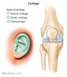
Types of Cartilage


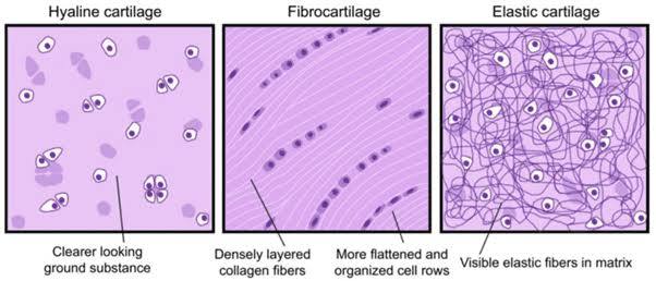
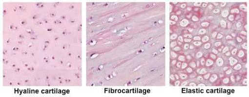
There are three main types of cartilage: Hyaline, Elastic, and Fibrocartilage, each with a distinct composition of collagen and elastin fibers, providing varied flexibility and strength for functions such as smooth joint movement, structural support in the ears/nose, and shock absorption in the spine. Hyaline is common and glassy, Elastic is flexible (ear, epiglottis), and Fibrocartilage is tough and strong (discs, menisci).
1. Hyaline Cartilage
- Appearance: Glassy (from Greek hyalos) due to fine, dispersed Type II collagen fibers, which are hard to see.
- Properties: Smooth, flexible, provides support, weakest type.
- Locations: Ends of long bones (articular cartilage), nose, ribs, trachea, larynx, embryonic skeleton.
2. Elastic Cartilage
- Appearance: Contains elastic fibers (elastin) and collagen, appearing yellowish.
- Properties: Strong and elastic, highly flexible.
- Locations: External ear, epiglottis, parts of the larynx.
3. Fibrocartilage
- Appearance: Dense, interwoven bundles of thick collagen fibers (Type I & II) within a hyaline matrix.
- Properties: Toughest, most durable, highly resistant to compression.
- Locations: Intervertebral discs, menisci (knee), pubic symphysis, tendon insertions.
Hyaline Cartilage
Hyaline cartilage is the glass-like (hyaline) and translucent cartilage found on many joint surfaces. It is also most commonly found in the ribs, nose, larynx, and trachea. Hyaline cartilage is pearl-gray in color, with a firm consistency, and has a considerable amount of collagen. It contains no nerves or blood vessels, and its structure is relatively simple.
Structure
Hyaline cartilage is the most common kind of cartilage in the human body. It is primarily composed of type II collagen and proteoglycans. Hyaline cartilage is located in the trachea, nose, epiphyseal plate, sternum, and ribs.
Hyaline cartilage is covered externally by a fibrous membrane known as the perichondrium. The primary cells of cartilage are chondrocytes, which are in a matrix of fibrous tissue, proteoglycans, and glycosaminoglycans.
As cartilage does not have lymph glands or blood vessels, the movements of solutes, including nutrients, occur via diffusion within the fluid compartments contiguous with adjacent tissues. Cartilage gives the structures a definite but pliable form, making them strong but with limited mobility and flexibility. Cartilage has no nerves.
Hyaline cartilage also forms the temporary embryonic skeleton, which is gradually replaced by bone, and the skeleton of elasmobranch fish.
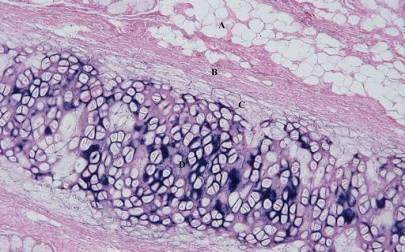
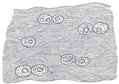
Microanatomy
When a slice of hyaline cartilage is examined under the microscope, it is shown to consist of chondrocytes of a rounded or bluntly angular form, lying in groups of two or more in a granular or almost homogeneous matrix. When arranged in groups of two or more, the chondrocytes have rounded, but generally straight outlines, where they are in contact with each other, and in the rest of their circumference, they are rounded.
They consist of translucent protoplasm with fine interlacing filaments, and minute granules are sometimes present. Embedded in this are one or two round nuclei, having the usual intranuclear network.
The cells are contained in cavities in the matrix, called cartilage lacunae. These cavities are actually artificial gaps formed from the shrinking of the cells during the staining and setting of the tissue for examination. The inter-territorial space between the isogenous cell groups contains relatively more collagen fibers, allowing it to maintain its shape while the actual cells shrink, creating the lacunae. This constitutes the so-called ‘capsule’ of the space. Each lacuna is usually occupied by a single cell, but during mitosis, it may contain two, four, or even eight cells.
Articular cartilage

Articular cartilage is hyaline cartilage on the articular surfaces of bones. It lies inside the joint cavity of synovial joints, bathed in synovial fluid produced by the synovial membrane, which lines the walls of the cavity.
Though it is often found in close contact with menisci and articular disks, articular cartilage is not considered a part of either of these structures, which are made entirely of fibrocartilage.
The articular cartilage extracellular matrix has a highly specialized architecture that is zonally organized: the superficial zone consists mostly of type II collagen fibers aligned parallel to the articular surface to resist shear forces, whereas the deep zone consists of the same fibers aligned perpendicularly to the bone interface to absorb compressive loads.
The biochemical breakdown of the articular cartilage results in osteoarthritis – the most common type of joint disease.[7] Osteoarthritis affects over 30 million individuals in the United States alone, and is the leading cause of chronic disability amongst the elderly.
Articular cartilage development begins with interzone condensation of a type II collagen-positive limb bud at the future joint site. This is followed by a definition of specific cellular subtypes (meniscal progenitors, articular progenitors, synovial progenitors, and ligament progenitors) that will eventually form the joint capsule. Finally, the joint capsule matures and forms a cavity, with a central meniscus, and an encasement of synovium.[9] This final structure will form several distinct layers of the articular cartilage found in all synovial joints, including the deep zone (closest to the bone), middle zone, and superficial zone (closest to the synovial fluid).
Maintenance of articular cartilage is guided by a balance of anabolic (cartilage-generating) and catabolic (cartilage degrading factors, like the maintenance of bone. Over the lifetime of the organism, anabolic factors and catabolic factors are generally in balance; however, as the organism ages, catabolism predominates, and cartilage begins to degrade. Eventually, the loss of hyaline cartilage matrix and reduction in the chondrocyte content of the hyaline cartilage matrix result in the development of joint disease such as osteoarthritis. Overexpression of hyaline-cartilage specific anabolic factors, such as FGF18, appears to restore the balance between cartilage loss and generation.
Elastic Cartilage


Elastic cartilage is a flexible connective tissue providing shape and support to structures needing to bend and snap back, like the outer ear (auricle), epiglottis, and larynx, due to its matrix rich in elastic fibers in addition to collagen type II. This tissue is yellow, surrounded by a perichondrium, and contains cells (chondrocytes) in lacunae, allowing for elasticity and strength.
Key Characteristics:
- Composition: Abundant network of elastic fibers, collagen type II, and proteoglycans within its matrix.
- Appearance: Typically yellowish in fresh specimens due to the elastic fibers.
- Structure: Chondrocytes (cartilage cells) reside in small cavities called lacunae, and it’s covered by perichondrium.
- Properties: Strong, flexible, and can return to its original shape after being deformed.
Locations:
- External Ear (Auricle/Pinna): Allows the ear to bend and move.
- Epiglottis: Cover the windpipe during swallowing.
- Larynx: Provides flexible support to vocal structures.
- Eustachian Tube (Auditory Tube): Part of the middle ear canal.
Function:
- Provides flexible support and maintains the shape of structures that undergo frequent bending and deformation, preventing damage.
Fibrocartilage


Fibrocartilage is the toughest type of cartilage, a hybrid tissue blending hyaline cartilage with dense fibrous connective tissue, characterized by thick bundles of Type I collagen fibers within a cartilage matrix, making it incredibly strong and shock-absorbent. It’s found in areas needing extreme durability and resistance to compression and tension, like intervertebral discs, knee menisci, pubic symphysis, and tendon/ligament insertions, acting as a shock absorber and connecting tissues.
Structure & Composition
- Collagen: Rich in dense Type I collagen fibers, organized into thick bundles, providing tensile strength.
- Cells: Contains both chondrocytes (cartilage cells) and fibroblasts (fibrous tissue cells).
- Matrix: A blend of cartilage ground substance and collagen fibers, allowing for toughness and some elasticity.
- No Perichondrium: Lacks a surrounding perichondrium (protective layer).
Key Functions
- Shock Absorption: Cushions bones and absorbs impact.
- Withstands Stress: Resists high compressive, tensile, and shear forces.
- Connects Tissues: Attaches tendons and ligaments to bone (entheses).
Locations in the Body
- Intervertebral Discs: Between vertebrae.
- Knee Menisci: In the knee joint.
- Pubic Symphysis: Where the two pubic bones meet.
- Tendon/Ligament Attachments: Sites where they join bone.
- TMJ (Temporomandibular Joint).
